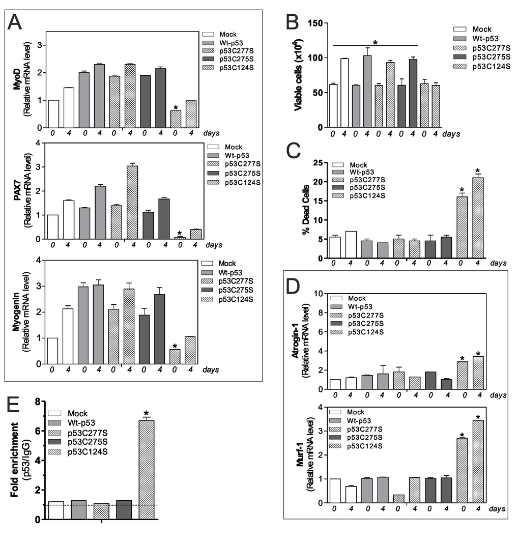
Figure 4.p53C124S mutant induces atrophy of C2C12 myoblasts. (A) C2C12 myoblasts were transfected with pcDNA3.1 vector containing cDNA for wild type p53 (Wt-p53), three single p53 mutants in DBD (p53C277S, p53C275S, p53C124S) or with empty vector (Mock). After 24 h from transfection C2C12 cells were differentiated for 4 days. Total RNA was isolated and relative mRNA levels of MyoD, PAX7 and Myogenin were analyzed by RT-qPCR. Data are expressed as means ± S.D. All the values were significantly different with respect to Mock day 0/4 (n=3, p<0.05; *p<0.05 was significantly decreased with respect Mock day 0). (B) Cells were counted by Trypan Blue exclusion. Data are expressed as means ± S.D. All the values were significantly different with respect to day 0 (n=4 *p<0.05). (C) Dead cells were counted by Trypan blue exclusion. Data are expressed as means ± SD (n=4, *p<0.001 vs Mock-, Wt-p53-, p53C277S- and p53C275S-day 0/4 cells). (D) Total RNA was isolated and relative mRNA levels of MuRF-1 and Atrogin-1 were analyzed by RT-qPCR. Data are expressed as means ± S.D. (n=3; *p<0.05 vs Mock-, Wt-p53-, p53C277S- and p53C275S-day 0/4 cells). (E) ChIP assay was carried out on cross-linked nuclei from Mock, Wt-p53, p53C277S, p53C275S and p53C124S cells at day 4 of myogenesis using p53 antibody followed by qPCR analysis of p53RE. Dashed line indicates the value of IgG control. Data are expressed as means ± S.D. (n=3; *p<0.001 vs Mock-, Wt-p53-, p53C277S- and p53C275S-day 4 cells).