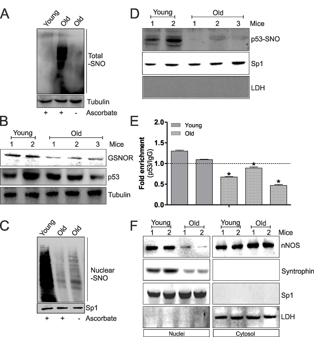
Figure 5.The decrement of nNOS nuclear localization inhibits p53 S-nitrosylation and its binding on ppargc1a promoter during aging. (A) Skeletal muscle of young (12 weeks) and old (80 weeks) mice was homogenized and total proteins (500 μg) were subjected to S-NO derivatization with biotin. After Western blot, biotin adducts were identified by incubating nitrocellulose membrane with HRP-conjugate streptavidin. Proteins incubated in labeling buffer without ascorbate were used as negative control (-Ascorbate). Tubulin was used as loading control. (B) Skeletal muscle of two young (12 weeks) and three old (80 weeks) mice was homogenized and total proteins (20 μg) were loaded for Western blot analysis of GSNOR and p53. Tubulin was used as loading control. (C) Skeletal muscle of young (12 weeks) and old (80 weeks) mice was homogenized and nuclear proteins (500 μg) were subjected to S-NO derivatization with biotin. After Western blot, biotin adducts were identified by incubating nitrocellulose membrane with HRP-conjugate streptavidin. Proteins incubated in labeling buffer without ascorbate were used as negative control (-Ascorbate). Sp1 was used as loading control. (D) Skeletal muscle of two young (12 weeks) and three old (80 weeks) mice was homogenized and nuclear proteins (500 μg) were subjected to S-NO derivatization with biotin. After Western blot the nitrocellulose was incubated with p53 antibody for detection of p53-SNO. Sp1 was used as loading control. The possible presence of cytoplasmic contaminants was tested by incubating nitrocellulose with rabbit anti-LDH. (E) ChIP assay was carried out on cross-linked nuclei from two young (12 weeks) and three old (80 weeks) mice using p53 antibody followed by qPCR analysis of p53RE. Dashed line indicates the value of IgG control. Data are expressed as means ± S.D. (n=3; *p<0.05). (F) Skeletal muscle of two young (12 weeks) and two old (80 weeks) mice was homogenized and 20 μg of nuclear and cytoplasmic extracts were loaded for detection of nNOS and Syntrophin by Western blot. Sp1 was used as loading control. The possible presence of cytoplasmic contaminants was tested by incubating nitrocellulose with rabbit anti-LDH. All the immunoblots reported are from one experiment representative of four that gave similar results.