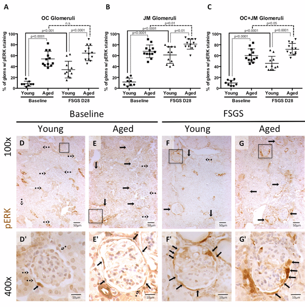
Figure 5.Percentage of glomeruli with phosphorylated-ERK stained PECs was highest in aged baseline mice with FSGS. (A-C) Quantitation showing the percentage of glomeruli with pERK staining of PECs along Bowman’s capsule. Aged mice and aged mice with FSGS had a higher percentages of glomeruli with pERK staining along Bowman’s capsule when compared to their respective young baseline and young FSGS mice in outer cortical glomeruli (OC) (A), juxta-medullary glomeruli (B) and combined OC and JM glomeruli(C). Overall, aged FSGS mice had the highest percentage of glomerular with pERK staining (C). (D-G) Representative images of pERK staining along Bowman’s capsule. Representative images of glomeruli at 100x magnification, with 400x magnifications shown in D’-G’ of the glomeruli marked by solid black square. Dashed arrows indicate pERK negative and solid arrows indicated pERK positive glomeruli (100x) and PECs (400x).