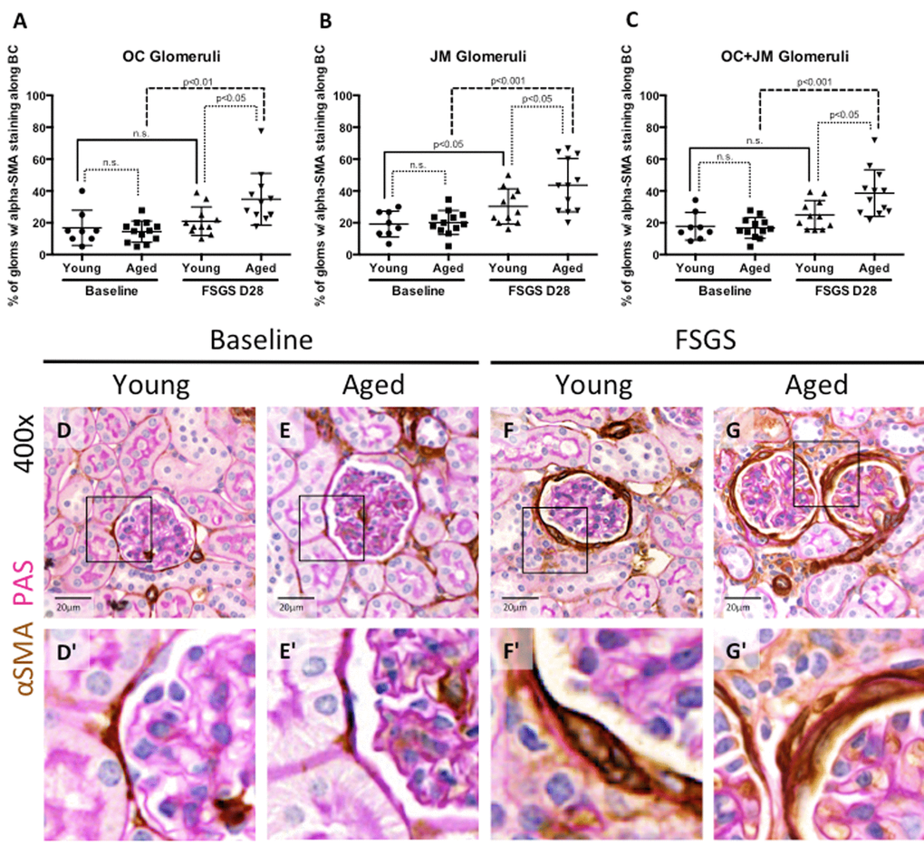
Figure 6.EMT marker staining along Bowman’s capsule was highest in aged FSGS mice. (A-C) Quantitation showing the percentage of glomeruli with α-SMA (EMT marker) staining along BC. There was no significant difference in young and aged mice at baseline in OC (A), JM (B), or combined (C) glomeruli. In OC (A), JM (B), and combined (C) glomeruli, α-SMA staining increased with disease in aged animals, while only in the JM (B) was α-SMA staining significantly increased in young mice, likely due to large variation within the sample groups. (D-G) Representative images of glomeruli with alpha-SMA staining along BC taken at 40x. Frequency and intensity α-SMA staining increased with disease (D vs. F, E vs. G), despite similar levels between young and aged animals at baseline (D vs. E). (D’-G’) Higher power images of the area demarcated by the solid square shown above, emphasizing the increase in α-SMA staining of cells along BC (F’, G’).