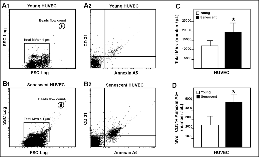
Figure 2.HUVEC-derived MVs assessed by flow cytometry. Representative dot plots showing log forward scatter (FSC) vs. log side scatter (SSC) from young (A1) and senescent (B1) HUVEC. Representative dot plots showing annexin A5 and CD31 phenotype from young (A2) and senescent (B2) HUVEC. (C) Absolute number of MVs per µL of sample from young and senescent HUVEC; *p<0.05. (D) Absolute number of CD31+/annexin A5+ MVs per µL of sample; *p<0.05. Results are presented as the mean ± SD of 6 independent experiments.