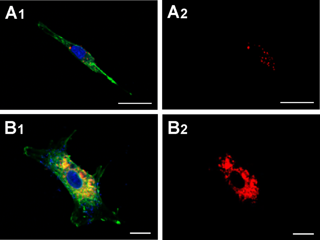
Figure 4.Capture of MVs by HASMC. Cells were incubated for 48 h with MVs from young (panels A1 and A2) or senescent HUVEC (panels B1 and B2) stained with CellTracker CM-Dil PI (red) and then were stained with phalloidin (PKH67, green). Nuclei were stained with DAPI (blue). Fluorescence was evaluated by confocal microscopy. (A) Scale bar 25µm, and (B) scale bar 10 µm. A representative experiment from three different experiments is shown.