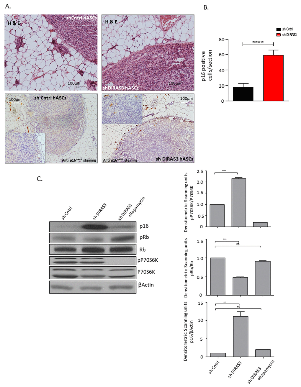
Figure 5.Human DIRAS3 KD ASCs develop a senescence phenotype in posterior sWAT of SCID mice. (A) (Upper panel) Human shDIRAS3 ASCs (shDIRAS3 hASCs) and shCntrl hASCs were xenotransplanted into posterior sWAT of SCID mice. Injection sites of hASCs were histologically identified and marked by H&E staining. (Lower panel) Senescent DIRAS3 KD hASCs were detected by immunohistochemical staining using anti p16INK4A antibodies. Region of Interest (ROI) is shown in higher magnification. (B) Number of p16INK4A positive hASCs per section were counted and plotted (n = 5 per group). (C) (Left panel) Cell lysates from control ASCs and DIRAS3 KD ASCs cultured with and without 20 nM rapamycin were blotted for p16INK4A, Rb, pRb (S807/811), P70S6K and pP70S6K (T389) using specific antibodies. β-Actin served as loading control. Note, rapamycin was added 2 days after virus infection. (Right panels) Fold changes in densitometric band intensities presented as Arbitrary Units (AU) for phosphorylated proteins normalized to un-phosphorylated total proteins, acquired by image J were compared. Band intensity of shCntrl was taken as 1 (n=2). All error bars represents the means ± SEM. p values * = p<0.05, **= p<0.001 and *** = p<.0001.