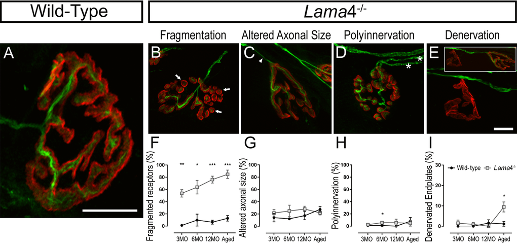
Figure 5.Accelerated aging features at lama4-/- neuromuscular junctions in EDL muscle. (A-E) Representative staining of axons and nerve terminal (green) in relation to the postsynaptic endplates (red) in WT (A) and lama4-/- (B-E) at 3MO. WT (A) showed normal innervation pattern while lama4-/- NMJs had (B) fragmented islands of postsynaptic receptors (as indicated by arrows), (C) altered innervating axonal size with thinning (as indicated by arrowhead) with some endplates displaying swollen axons, (D) polyinnervation or multiple axons entering the nerve terminal (as indicated with asterisks) and (E) completely denervated endplate with a smaller insert panel showing an occupied endplate alongside the denervated endplate. Scale bar = 10 µm. Quantification of aging features; (F) fragmented endplates, (G) altered axonal size, (H) polyinnervation and (I) denervated endplates across all ages. (F-I) n = 3, NMJs = 63-109 per genotype at each age group. Statistical analyses were performed using Student’s t-tests; values are presented as mean ± SEM (* P < 0.05, ** P < 0.01, and ***P < 0.001).