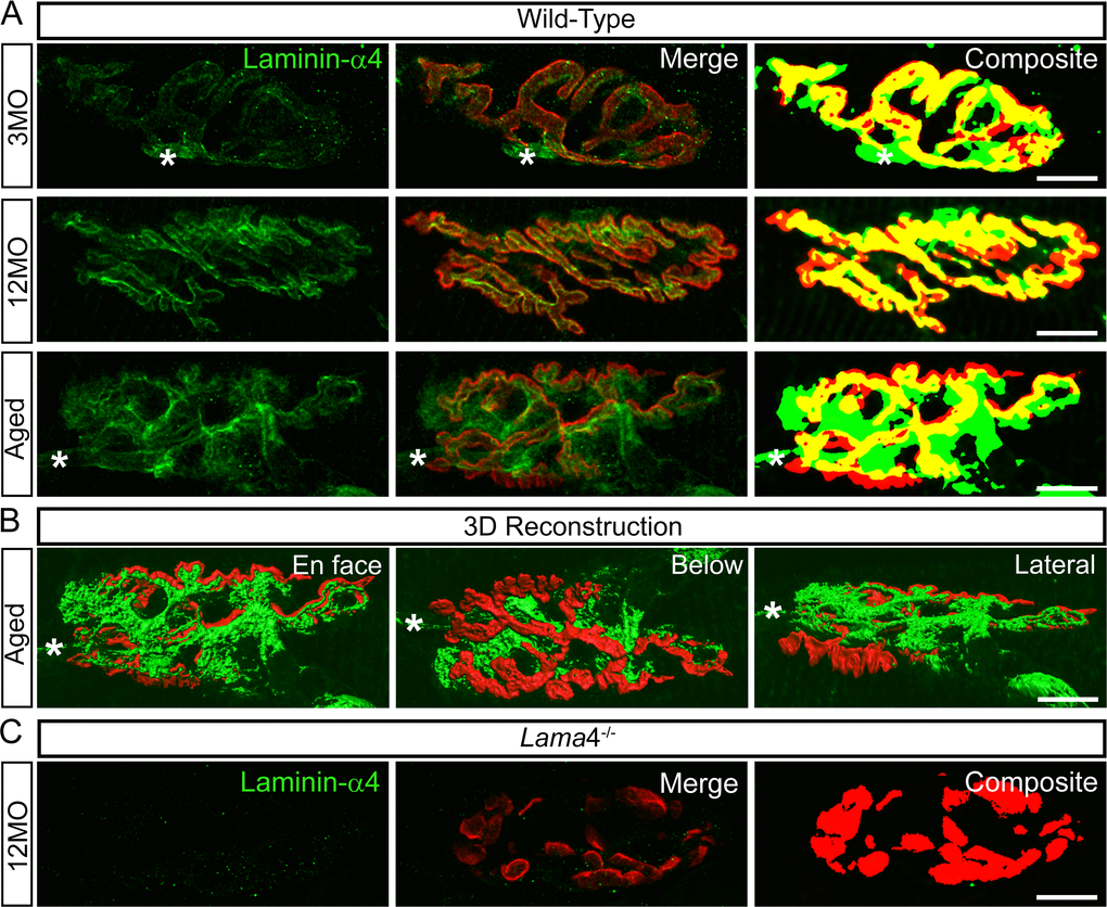
Figure 7.Altered expression of laminin-α4 at 12MO wild-type NMJs, becoming more evident at aged neuromuscular junctions. (A) Examples of representative staining of laminin-α4 chain (green) with respect to the postsynaptic endplates (red) in EDL muscle. Composite panel showed colocalization of laminin-α4 with postsynaptic endplate = yellow, extrasynaptic localization of laminin-α4 = green and missing laminin-α4 expression at postsynaptic region = red. Asterisk (*) is an indication of a nerve entering the nerve terminal, therefore it is not considered as an extrasynaptic localization of the laminin-α4 chain. WT at 3MO showed normal colocalization of laminin-α4 chain with respect to the postsynaptic endplates. By 12MO, WT began to display missing expression of laminin-α4 (observed with more red on composite panel) and mislocalized laminin-α4 expression (more green) compared with 3MO WT. As the NMJ aged, WT showed drastic changes in laminin-α4 expression with highly expressed laminin-α4 at the extrasynaptic region (high degree of green) and lacking laminin-α4 (high degree of red) compared with 12MO WT. The overlapping of laminin-α4 to postsynaptic endplates (yellow) at aged WT NMJ was at a much lesser degree in comparison with 12MO WT. (B) 3-dimensional (3D) reconstruction of laminin-α4 staining (green) in relation to postsynaptic endplate (red) of aged WT NMJ from (A), in three different planes of view; en face, below and lateral. En face view is reflective of the image shown in (A). Below and lateral views confirmed that non-colocalized laminin-α4 staining was purely expressed at the extrasynaptic basal lamina region, not by non-muscle cells. (C) Lama4-/- NMJs at 12MO displayed no expression of laminin-α4 chain. Scale bar = 10 µm.