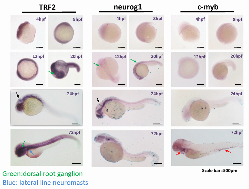
Figure 5.TERFA expression increase since neural development at embryonic stage and remains high in the brain during larval stage development. Representative photomicrographs of whole-mount in situ hybridization of TERFA, Neurog1 and c-MYB mRNA. The RNA probe labelled with DIG was stained in dark blue. The green arrow indicates the dorsal root ganglion neuron and the blue arrow indicates the lateral line neuromasts. The black arrow in 24hpf indicates the midbrain boundary. The red arrow indicates the c-MYB signal marked hematopoietic tissue.