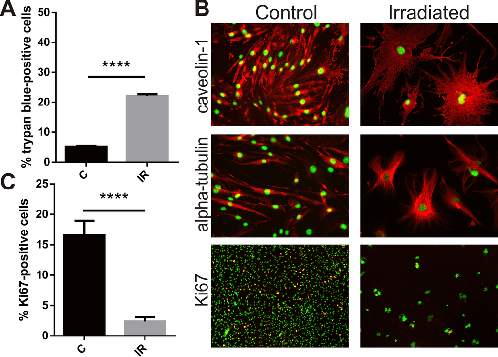
Figure 1.Radiation inhibits proliferation, induces cell death and hypertrophy in brain endothelial cells. Mouse bEnd.3 cells were delivered a dose of 20 Gy ionizing radiation by linear accelerator. (A) Floating and adherent cells were collected at day 6 and cell viability/death measured by trypan blue staining and counting in a Neubauer chamber. Data show trypan blue positive cells as a percentage of total cells. Mean of 3 independent experiments ± SEM. (B) Representative images of non-irradiated (control) and irradiated bEnd.3 cells stained after permeabilization for caveolin-1 or α-tubulin showing cell hypertrophy and cytoskeletal rearrangement at day 6 (red, 200× magnification), or proliferation marker Ki67 (lower panels, red, 100× magnification). Nuclei were counterstained with DAPI (green). Co-localization (yellow). (C) The proportion of Ki67 positive cells was determined using Image J. Data represent mean ± SEM calculated in 5 fields of view (100×) from 2 independent experiments. Student’s t-test, ****P<0.0001.