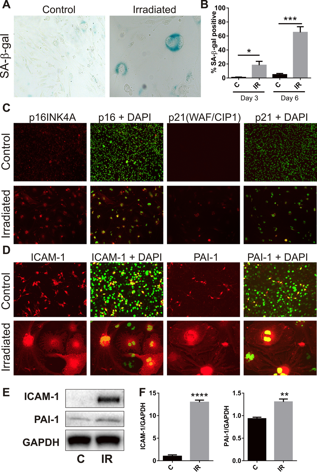
Figure 2.Radiation induces senescence-associated markers. (A) Representative bright field images of non-irradiated (control) and irradiated bEnd.3 cells stained for lysosomal SA-β-gal activity (perinuclear blue staining) at day 6 post-IR or sham (100× magnification). (B) The proportion of SA-β-gal-positive cells was quantitated at day 3 and day 6 in 3–4 independent experiments. (C) Representative immunofluorescent images of nuclear accumulation of CDK inhibitors, p21 and p16 (red), in irradiated cells after 6 days (red, 100× magnification). (D) Representative immunofluorescent images of ICAM-1 and PAI-1 staining in control and irradiated cells after 6 days (red, 200× magnification). Cells were counterstained with cell surface marker wheat germ agglutinin conjugated to AF488 (blue). Cell nuclei were stained with DAPI in all merged images (green). (E) ICAM-1 and PAI-1 expression were determined in control and irradiated cells by western blotting and quantitated after normalization to GAPDH using Image J (Figure F; n=4 independent experiments). All data are shown as mean ± SEM. Student’s t-test *P<0.05, *** P<0.01, ***P<0.001, ****p<0.0001. C, control; IR, irradiated.