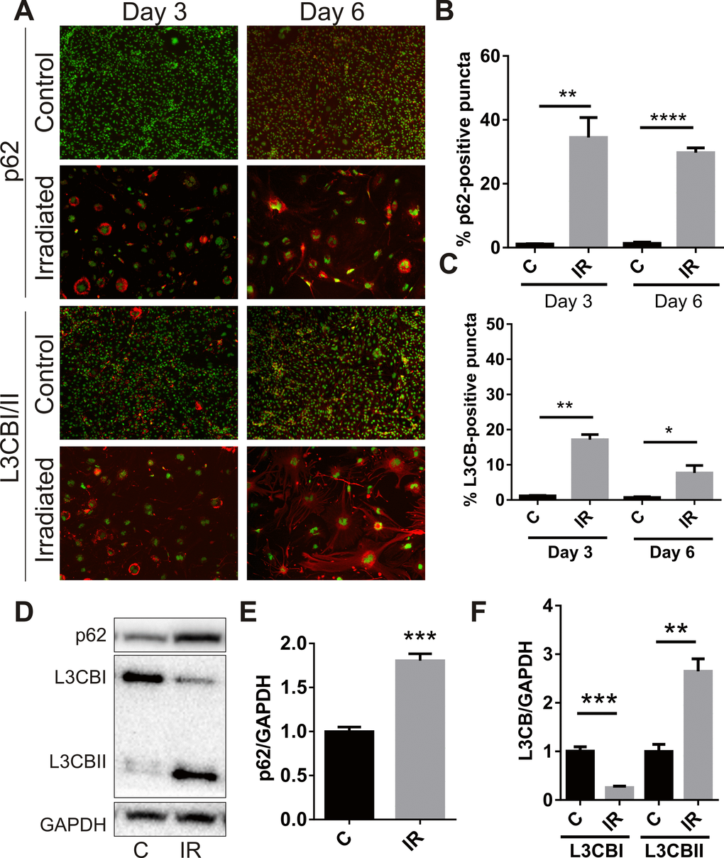
Figure 3.Radiation stimulates accumulation of autophagy-associated markers in brain endothelial cells. bEnd.3 cells were delivered a dose of 20 Gy ionizing radiation and monitored for 3–6 days. (A) Representative immunofluorescent images showing increased perinuclear p62 or L3CB-positive puncta accumulating in permeabilized cells at day 3 and day 6 post-IR or sham (red, 100× magnification). Cells were counterstained with DAPI (green). The percentage of cells positive for p62 (B) or L3CBI/II puncta (C) were quantitated using Image J (n=3 independent experiments; positive cells were counted in n=8 fields of view). (D) Representative western blots of p62, L3CBI and II autophagosomal markers in whole cell lysates of control and irradiated cells after 6 days. (E) and (F) Bands were quantitated after normalization to GAPDH using Image J (n=4 independent experiments). All data are shown as mean ± SEM. Student’s t-test, *P<0.05, *** P<0.01, ***P<0.001, ****p<0.0001. C, control; IR, irradiated.