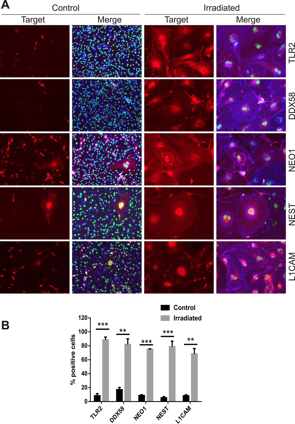
Figure 5.Immunofluorescent localization of ADAM10 target proteins. (A) Representative immunofluorescent images of non-permeabilized cells stained for L1CAM, NEST, NEO1, DDX58 and TLR2 after 6 days post-IR or sham (controls). Cells were co-stained with DAPI (nuclei, green) and wheat germ agglutinin–AF488 (surface marker, blue). All images are shown at 200× magnification. (B) Percentage of cells staining positively for each target protein 6 days after IR or sham. Data represent at least 3 independent experiments, positive cells were counted in n=3 fields of view. All data shown as mean ± SEM. Student’s t-test **P<0.01, ***P<0.001. C, control; IR, irradiated.