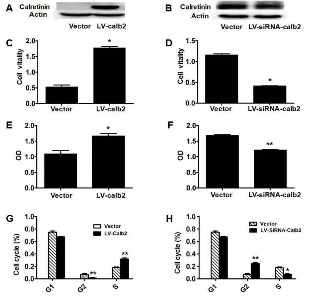
Figure 1.Effect of calretinin on Leydig cell proliferation. (A) Calretinin expression in MLTC-1 cells transfected with LV-calb2. The vector was used as the negative control. (B) Calretinin expression in R2C cells transfected with LV-siRNA-calb2 or vector (as control). (C-H) After MLTC-1 and R2C cells were transfected with LV-calb2, LV-siRNA-calb2 and vector (as negative control), cell viability was detected using CCK8 kits, cell proliferation was tested with BrdU proliferation assay kits and cell cycle position was analyzed by flow cytometry. (C) The viability of MLTC-1 cells with up-regulated calretinin was significantly higher when compared with the control group. (D) The viability of R2C cells with down-regulated calretinin was significantly lower. (E) The OD of MLTC-1 cells with up-regulated calretinin was significantly higher compared with the control group. (F) The OD of R2C cells with down-regulated calretinin was significantly decreased. (G) When calretinin was up-regulated in MLTC-1 cells, the number of cells in the G2 phase significantly decreased while the number of S phase cells significantly increased. (H) When calretinin was down-regulated in the R2C cells, the number of cells in the G2 phase significantly increased while the number of S phase cells significantly decreased. * p<0.05; **: p<0.01.