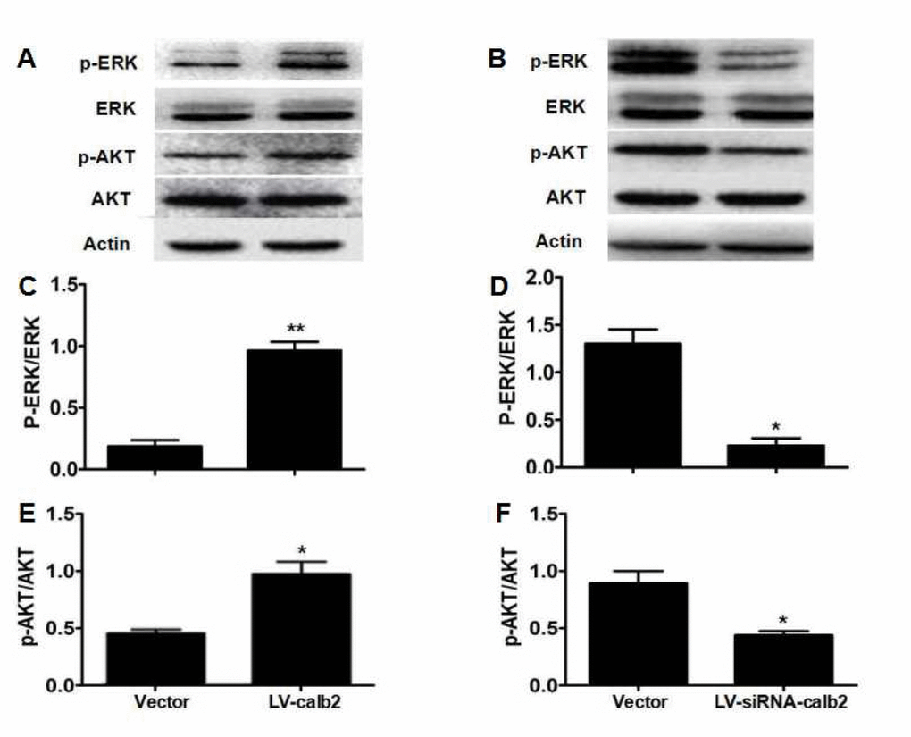
Figure 2.Regulation of calretinin on Leydig cell proliferation partially via the ERK1/2 and AKT pathways. (A) AKT, p-AKT, ERK1/2 and p-ERK1/2 expression in MLTC-1 cells with over-expressed calretinin. (B) AKT, p-AKT, ERK1/2 and p-ERK1/2 expression in R2C cells with down-regulated calretinin. (C) The ratio of p-ERK1/2 /total ERK1/2 was significantly higher when calretinin was up-regulated in MLTC-1 cells. (D) The ratio of p-ERK1/2/total ERK1/2 was significantly lower when calretinin was down-regulated in R2C cells. (E) In MLTC-1 cells with up-regulated calretinin, the ratio of p-AKT/total AKT was significantly higher. (F) In R2C cells with down-regulated calretinin, the ratio of p-AKT/total AKT was significantly lower. The vector was used as the negative control(s). * p<0.05; **: p<0.01.