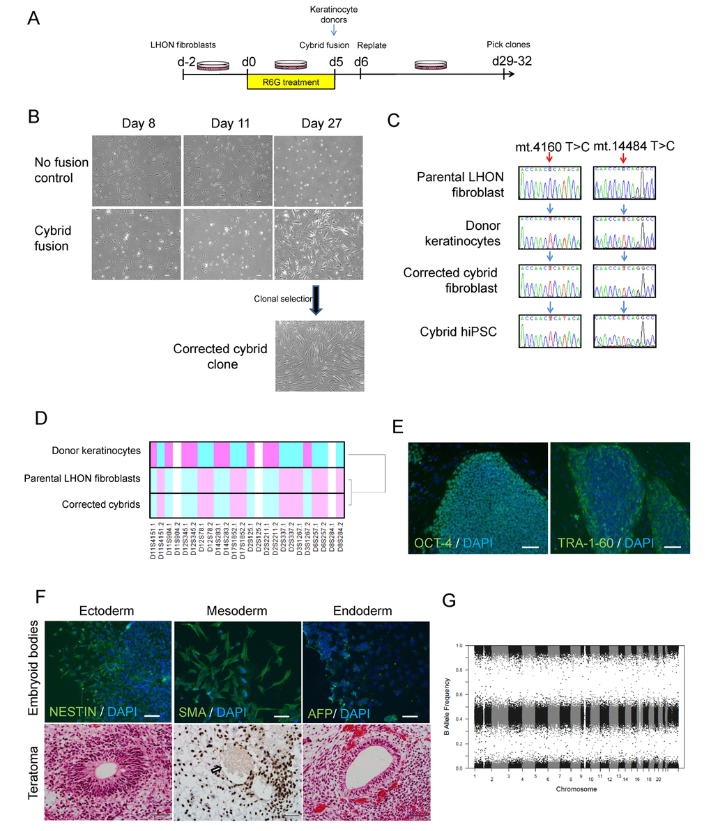
Figure 1.Using cybrid transfer to generate mutation-free LHON fibroblasts and iPSCs. (A) Diagram of cybrid generation. Fibroblasts were pre-treated with the mitochondrial toxin rhodamine 6-G (R6G) then fused with healthy donor mitochondria obtained from normal keratinocytes. On day 29-32, proliferating colonies were picked and expanded. (B) Representative images of control fibroblasts (no fusion) and fused fibroblasts that received donor mitochondria (cybrid fusion) at 8, 11, 27 days post R6G treatment. (C) Genotype confirming cybrid correction of mutation in fibroblasts and the corresponding iPSCs. Red arrows indicate LHON mutations at m.4160T>C and m.14484T>C, blue arrows indicate wild-type genotype. Note that the genotype of the parental LHON fibroblasts (LHON Q1-4) was reported previously [12]. (D) Microsatellite analysis confirming cybrid originated from LHON fibroblasts, but not donor keratinocytes. (E-G) Characterization of cybrid iPSCs (CYB iPSC c1). (E) Immunostaining showed expression of the pluripotency markers OCT-4 and TRA-1-60 in cybrid iPSCs. Scale bars: 100 μm. (F) Top panel: Differentiation of cybrid iPSCs by embryoid body formation contained cells positive for NESTIN (ectoderm), SMA (mesoderm) and AFP (endoderm) expression. Cells were counterstained with DAPI (blue). Scale bars: 100 μm. Bottom panel: Teratoma formation upon transplantation of cybrid iPSCs in nude rats, showing differentiation to endoderm, mesoderm (Ku80 staining, the arrow indicates an endothelial-lined blood vessel with lumen filled with red blood cells) and ectoderm. Scale bars: 50 μm. (G) Copy number variation analysis showing normal karyotype in cybrid iPSCs.