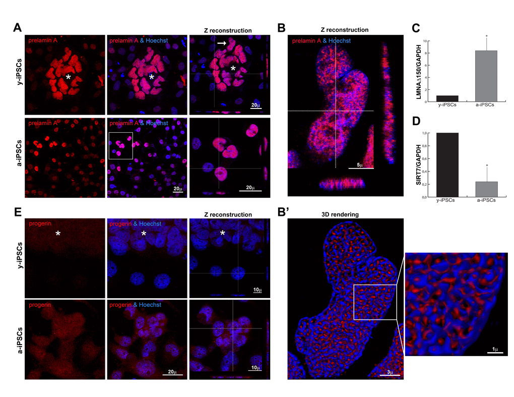
Figure 2.Distribution of prelamin isoforms and expression of LMNAΔ150 and SIRT7 in iPSCs, in pluripotent state and following differentiation and senescence. (A) Prelamin A was throughly distributed inside iPSCs nuclei of the colony cells (asterisk) and aged cells, as represented by XYZ stacks (right column), and it colocalized with nucleic acid stain (Hoechst, blue), as visualized in high magnifications (of the inset for a-iPSCs, and of y-iPSCs nuclei (arrow) showed in (B). (B) XYZ high magnification of y-iPSCs nuclei (indicated in a, arrow) showing a clear intranuclear accumulation of prelamin A in pluripotent stem cells. (B’) Deconvolved 3D-rendering of raw Z stack (in B) showing tight interconnections between prelamin A and chromatin distribution (high magnification of the inset, right). (C) Relative expression levels of LMNAΔ150 mRNA expression in y-iPSCs (*: p < 0,05, n=3). (D) Relative expression levels of SIRT7 mRNA in y-iPSCs and a-iPSCs (*: p <0,05, n=3). (E) Progerin expression was undetectable in y-iPSCs of the colony (asterisk) and in differentiated cells, whereas it appeared in senescent iPSCs.