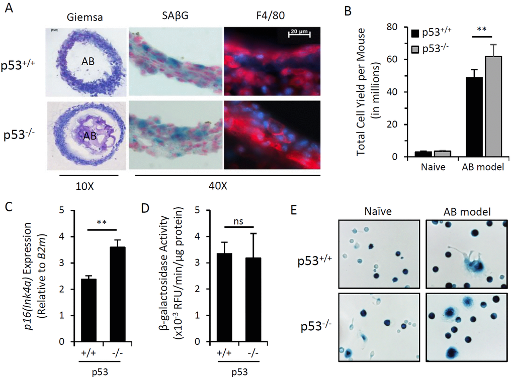
Figure 1.Induction of p16Ink4a and SAβG in macrophages does not require p53. Peritoneal lavage and alginate beads containing SCs (AB) were recovered from wild type (p53+/+) or p53 knockout (p53-/-) mice 15 days after injection (AB model). (A) Representative microphotographs of cryosectioned immunocyte capsules surrounding alginate beads stained with May-Grünwald-Giemsa for histology (10x objective), X-Gal substrate for β-galactosidase activity (SAβG; pH 6.0) (blue) with nuclear fast red counterstain (red), and an immunofluorescent antibody against macrophage marker F4/80 (red) with DAPI nuclear counterstain (blue) (40x objective). (B) Total yield of cells recovered from peritoneal lavage from naïve or AB-injected p53+/+ and p53-/- mouse strains. (C) AB model-elicited immunocyte capsules were pooled equally from 3 mice and p16Ink4a gene expression relative to internal reference gene β2-microglobulin (B2m) was measured by qPCR. (D) β-galactosidase activity from cell extracts of immunocyte capsules from alginate beads recovered from individual mice was measured via 4-MUG hydrolysis, presented as the rate of 4-MU fluorescence (RFU) per minute normalized per microgram of protein. (E) Representative microphotograph of adherence-selected peritoneal lavage from naïve and AB-injected mice stained with X-Gal for SAβG activity. Data show mean ± standard deviation of two independent experiments (n=3 mice per experiment). Statistical comparison between p53+/+ and p53-/- strains are indicated; ns, not significant; **, p-value < 0.01.