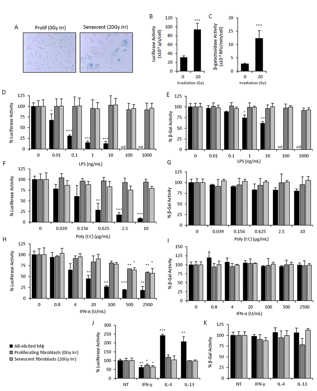
Figure 4.Elevated p16Ink4a and β-galactosidase is regulated by immunomodulatory agents in macrophages but not mesenchymal SCs. Primary cultures of adipose-derived mesenchymal stromal cells (AdMSC) isolated from p16Ink4a/Luc mice were irradiated (20Gy) and cultured for 10 days for senescence induction. Mock irradiated cells were passaged and used as a proliferating cell control. Response of senescent and proliferating AdMSCs to immunomodulatory agents were compared to that of peritoneal lavage cells elicited by the alginate bead model. (A-C) Characterization of senescent and proliferating AdMSCs. Microphotographs of SAβG-stained cells depicts positive staining of senescent cells, as well as an enlarged and flattened morphology, compared to that of proliferating cell control (A). p16Ink4a promoter-driven luciferase activity (B) and β-galactosidase activity measured via 4-MUG hydrolysis (C) were measured in senescent and proliferating AdMSCs, confirming senescent phenotypes. (D-K) Dose-response curves of LPS (D&E), Poly(I:C) (F&G), IFN-α (H&I), and IFNγ (10 ng/mL), IL-4 (20 ng/mL) and IL-13 (10 ng/mL) (J&K) on p16Ink4a promoter-driven luciferase activity (left panels: D,F,H& J) and β-galactosidase activity measured via 4-MUG hydrolysis (right panels: E,G,I&K) after 72hr treatment. No effect on viability was observed via CyQuant Direct assay (>80% viability). Results are shown as the mean ± standard deviation for at least 3 experiments, with statistical comparison to non-treated controls; *, p-value < 0.05; **, p-value < 0.01; ***, p-value < 0.001. nd, not determined.