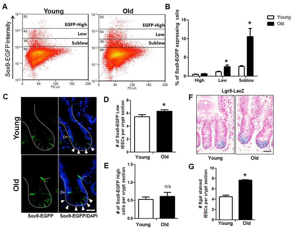
Figure 3.Increased IESC in old mice. (A) Representative flow cytometry data of Sox9-EGFP expressing cells in young and old Sox9-EGFP reporter mice. Gate: R3=Sox9-EGFPHigh, R4=Sox9-EGFPLow, R5=Sox9-EGFPSublow. (B) Relative abundance of different Sox9-EGFP expressing cells measured by flow cytometry. n=19 animals per group, *p<0.05 Young vs. Old, unpaired t test. (C) Representative images of crypt sections from young and old Sox9-EGFP reporter mice stained with EGFP and the nuclear marker DAPI. Sox9-EGFPLow IESC marked by closed triangles. Sox9-EGFPHigh EEC marked by open triangles. Magnification: 40x, Scale bar: 20µm. (D) Quantification of the number of Sox9-EGFPLow IESC counted per crypt section. n=8 young and 9 old animals, *p<0.05 Young vs. Old, unpaired t test. (E) Quantification of the number of Sox9-EGFPHigh cells counted per crypt section. n=6 young and 7 old animals. (F) Representative images of Xgal stained crypt sections from young and old Lgr5-LacZ reporter mice. Magnification: 40x, Scale bar: 20µm. (G) Quantification of the number of Xgal stained Lgr5-LacZ IESC counted per crypt section. n=4, p<0.05 Young vs. Old, unpaired t test.