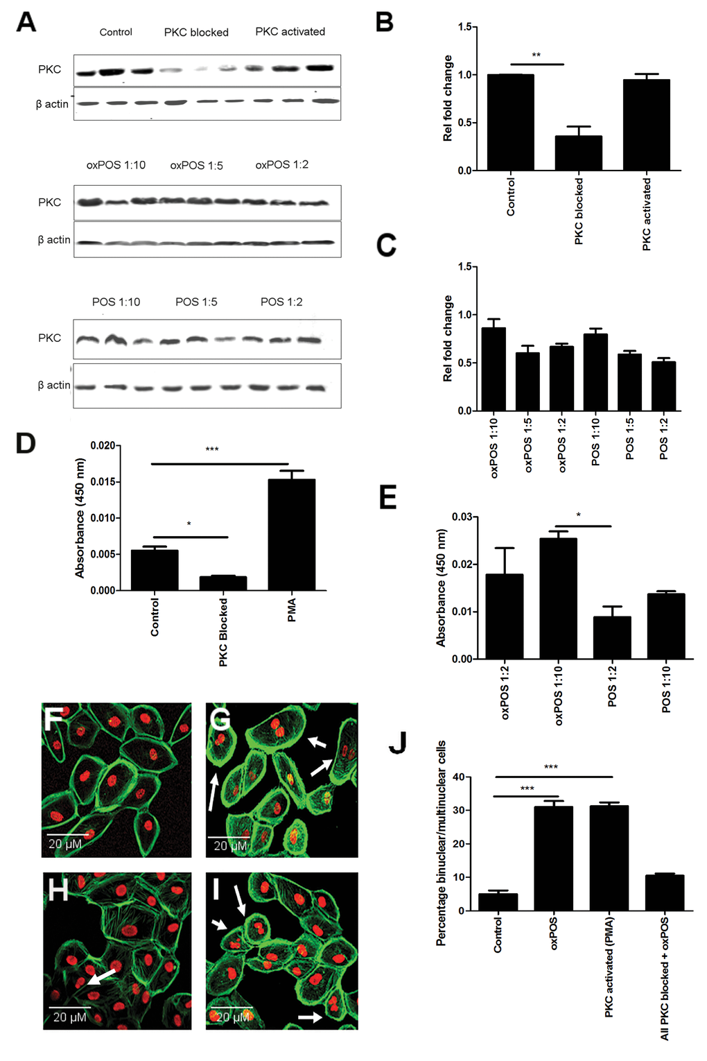
Figure 1.Role of PKC in RPE cell multinucleation and oxPOS induced PKC activation in RPE cells. (A) Representative Western blots showing total PKC protein expression in RPE cells following PMA, total PKC blockade, no treatment and with different concentrations of POS or oxPOS treatments (RPE: POS/oxPOS 1:2, 1:5, 1:10) for 48h. (B-C) quantification of total PKC expression in different treatment groups. The data are represented as relative fold change to the control. (D) Active PKC levels in RPE cells following PMA, total PKC blockade treatments, or untreated control for 48h. (E) Active PKC protein levels in RPE cells following POS or oxPOS treatment at 1:10 and 1:2 (RPE: POS/oxPOS) for 48h. Cells from each treatment group was homogenised and protein levels were evaluated by active PKC ELISA kit. *, P < 0.05; ***, P < 0.001 compared to the control untreated group. One-way ANOVA followed by Dunnett’s multiple comparison test. N = 3. (F-I): histochemical staining of propidium iodide (PI) and Phalloidin in control untreated ARPE19 (F), PMA treated ARPE19 (G), PKC blocked ARPE19 (H) and oxPOS (I) treated ARPE19 cells. Arrows indicate multinucleate RPE cells. (J) histogram showing the percentage of binucleated and multinucleate RPE cells, ***, P < 0.001 compared to control untreated. One-way ANOVA followed by Dunnett’s multiple comparison test. 50 cells were counted from three wells for each group.