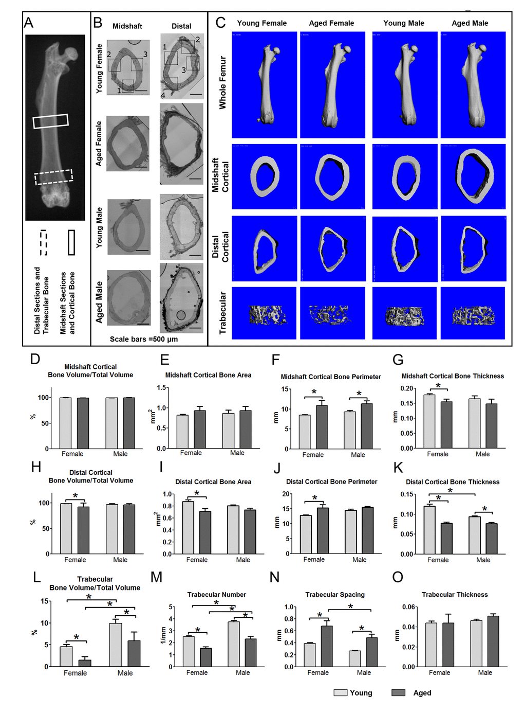
Figure 1.Aging is associated with structural changes in bone that are more pronounced in females than males. (A) X-ray of mouse femur showing regions used for midshaft cortical and distal cortical and trabecular analysis. (B) Bright field montaged images of midshaft and distal sections used for confocal imaging of 5 and 22mo mouse femurs with imaging regions indicated by numbered black boxes (Bars = 500μm). (C) MicroCT reconstructions for whole femurs and midshaft cortical and distal cortical and trabecular bone. Graphs in (D-G) show microCT quantitation of midshaft cortical bone parameters, (H-K) show distal cortical bone parameters and (L-O) show quantitation of trabecular bone parameters in young and aged mice. (Data are mean ± SEM, * = p< 0.05, ANOVA/Tukey’s, females n= 8, males n= 6-7) (Note: n=6 was used for the aged female group in graphs (M-O) because in two aged females the trabecular number was so low that the software cannot reliably compute values for trabecular spacing).