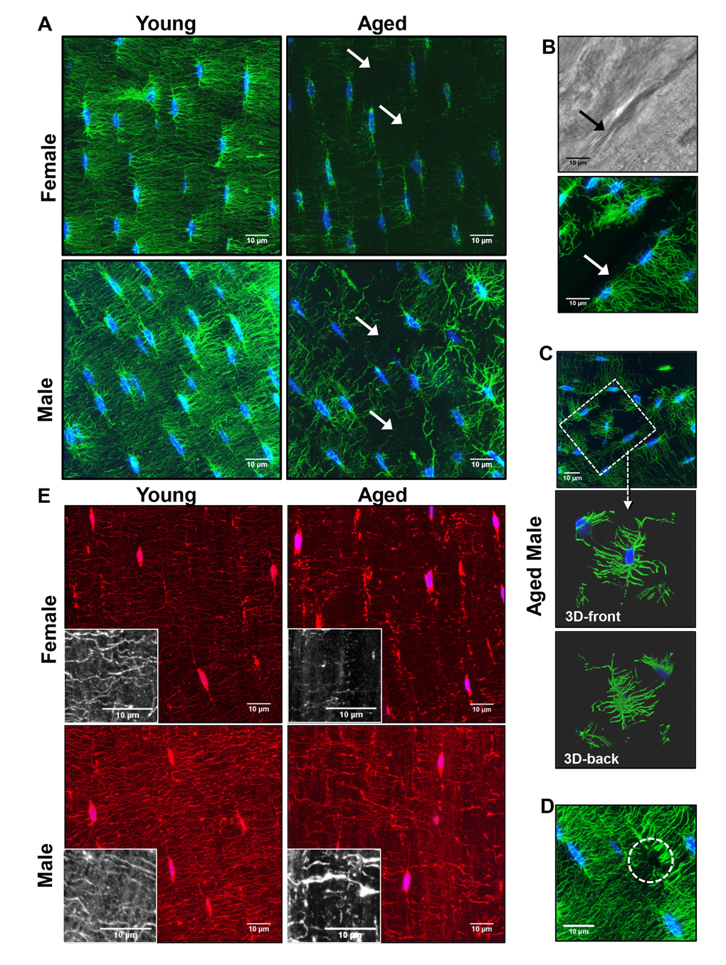
Figure 3.Degeneration of the osteocyte network in aged bone. (A) Maximal Z-projections of 250 planes (32.5μm) from 100x confocal images of phalloidin (green) and DAPI (blue) stained midshaft femur sections showing osteocyte connectivity in young and aged male and female mice. (B) Dendrites do not cross over discontinuities in the bone (arrow) although matrix is present as seen in the corresponding brightfield image. (C) Some osteocytes in aged animals are found in “islands” with few or no connections to the surrounding osteocytes (dashed box). Enlarged images in (C) show a 3D render (front and back) of the same osteocyte confirming its lack of connectivity with surrounding osteocytes. (D) Occasionally dendrites with no visible cell body were seen in the aged mice (dashed circle) suggesting the dendrites may be left behind after apoptosis. (E) Maximal Z-projections of 20 planes (2.6μm) from DiI (red) and DAPI (blue) labeled femur sections showing staining of the osteocyte cell membrane in young and aged mice. Black and white insets show lipid material in the matrix around the dendrites. (Bars = 10μm).