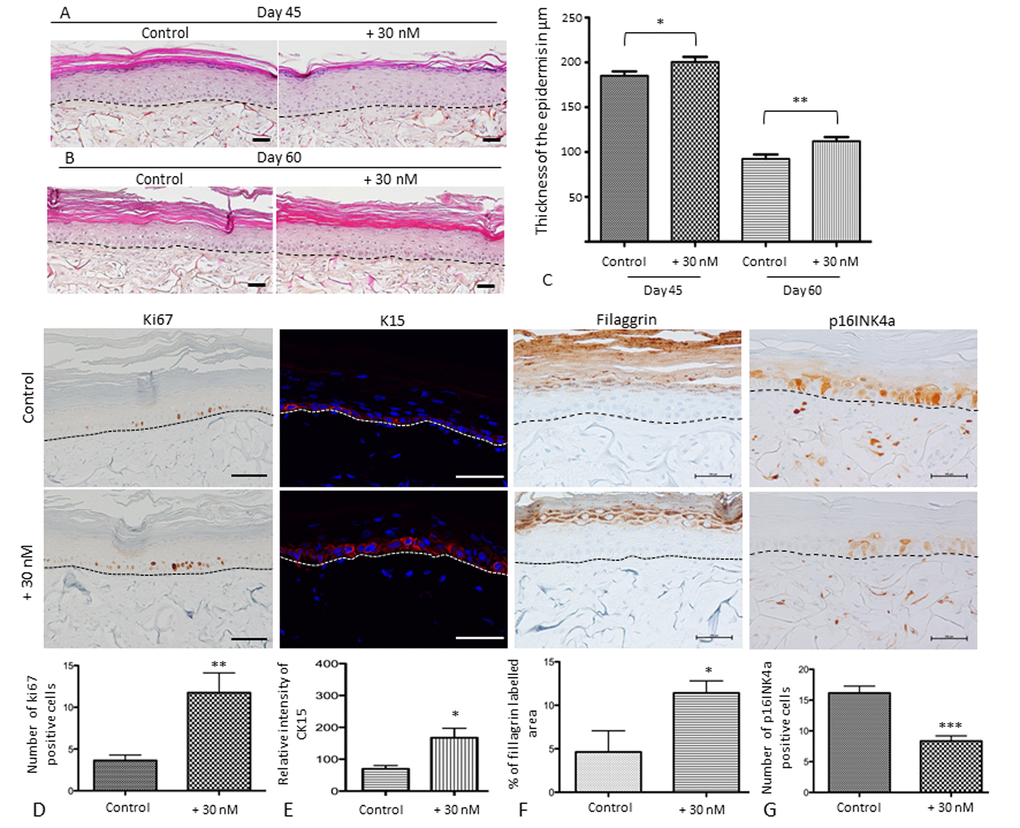
Figure 5.Sodium selenite supplementation improves skin equivalent quality during in vitro senescence and characterization of the balance between proliferation and differentiation of SEs during aging at day 60. Skin equivalents are generated from young keratinocyte and fibroblast donors and cultivated for 45 or 60 days to mimic skin aging [24]. The morphological aspects of SEs that were treated (+30 nM) or not (control) at (A) day 45 (D45) corresponding to 17 days of application and (B) day 60 (D60) corresponding to 32 days of application. Scale bar = 100 µm (C) Quantification of the thickness using ImageJ software is expressed in µm as a distance between from the basal layer of the epidermis to the stratum granulosum excepted for the stratum corneum. (D) Immunohistochemical staining of Ki67 in treated (+30 nM) or not (control) SEs and the average number of Ki67-expressing cells (scale bar = 500 µm). (E) Immunofluorescence staining of Cytokeratin 15 in treated (+30 nM) or not (control) SEs and the quantification of pixel intensity in relative units. (F) Immunohistochemical staining of filaggrin in SEs that were treated (+30 nM) or not (control) and quantification of labeled area in the living epidermis (scale bar = 500 µm). (G) Immunohistochemical staining of p16INK4a expression in SEs that were treated (+30 nM) or not (control) and quantification of the number of p16INK4a expressing cells (scale bar = 100 µm). The dermo-epidermal junction is indicated by a dotted line. Results are mean ± SD of 3 independent fields obtained from 3 independent samples. Representative photographs are shown. *p<0.05, **p<0.01.
Figure 5 — Selenium preserves keratinocyte stemness and delays senescence by maintaining epidermal adhesion | Aging