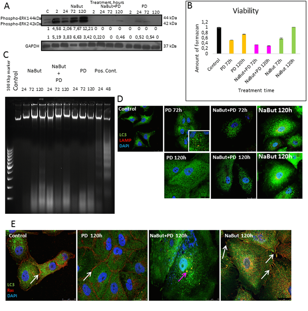
Figure 10.Senescent human Ki-Ras-mutated lung adenocarcinoma A549 cells after MEK/ERK suppression undergo autophagy impairment, Ras relocalization and cell death. (A) Western-blot analysis of ERK1,2 phosphorylation after short-term (2 h) and long-term (24 h-120 h) NaBut, NaBut + PD and PD treatment. Cells were exposed to inhibitors for the indicated time and then lysed and processed to Western-blotting in 12% gel. Numbers below present densitometry of bands. (B) Cell viability analyzed by MTT-test. In the indicated time intervals cells were processed with MTT reagent, the amount of formazan was measured at 570 nm wavelength. Data are presented as mean ±S.E.M. of three independent experiments (n=3). (C) Electrophoresis in 2% agarose gel of DNA extracted from untreated cells and cells after exposure to inhibitors shows the absence of nucleosomal DNA fragmentation in treated cells. DNA visualized with ethidium bromide. DNA extracted from serum-starved ERas cells undergoing apoptosis was used as a positive control. (D) Immunofluorescence images demonstrating LC3 and LAMP1 levels and colocalization after 72 h and 120 h of treatment. Cells were treated with inhibitors or left untreated and stained with antibodies against pan-LC3 (green) and LAMP1 (red). Nuclei stained with DAPI (blue). Scale bars: 25 µm. (E) Immunofluorescent images showing Ras (red) and LC3 (green) localization after 120 h of treatment. Cells were exposed to inhibitors for indicated time, then fixed and stained with antibodies against pan-Ras and pan-LC3. White arrows show membrane-bound Ras; pink arrow shows Ras in cytoplasm of senescent cells with inhibited MEK/ERK. Nuclei stained with DAPI (blue). Scale bars: 25 µm.