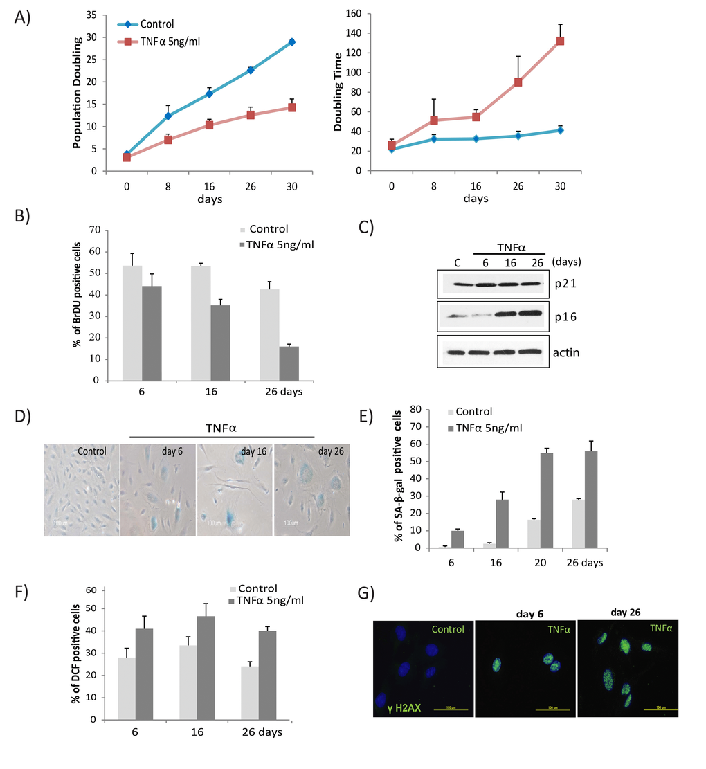
Figure 1.TNFα induces senescence and DNA damage in HUVECs. (A) Long-term growth curve of cells exposed to recombinant human TNFα (5ng/ml). Untreated cells were used as controls. Population doubling and doubling times were calculated based on cell density at confluence. Data represent mean values from 3 independent experiments. (B) The percentage of BrdU-positive cells was determined by FACS analysis in cells untreated or chronically treated with TNFα at the concentration indicated. (C) Western blot analysis of p21, p16, and actin in cells treated with TNFα 5ng/ml for the indicated times. (D) SA-β-gal activity in TNFα (5ng/ml)-treated or control cells for the indicated number of days. (E) Percentages of SA-β-gal-positive cells in control or TNFα-treated cultures. The data represent 2 independent counts of 200 cells from 3 independent experiments. (F) Intracellular ROS levels were monitored by 2',7’-dichlorodihydrofluorescein diacetate staining followed by flow cytometry. Bar graph represents percentage of DCFDA-positive cells treated with TNFα or medium alone. (G) Immunofluorescence detection of γH2AX foci in controls or cells treated with TNFα (5ng/ml) for indicated days. Data in A, B, E, and F represent mean value ± standard deviation (s.d.) from n=3, 2, 3, and 2 independent experiments, respectively.