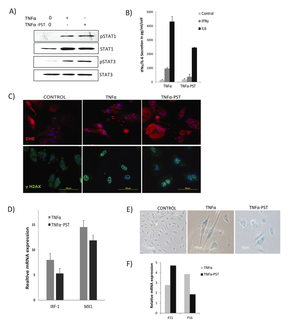
Figure 5.Persistent activation of STAT1/3. Cells were exposed to TNFα (20ng/ml) for 3 days, then washed to remove the residual TNFα and cultured for 3 days in the absence of exogenous TNFα (TNFα-PST). Parallel cultures were exposed to exogenous TNFα throughout the experiment. (A) Levels of p-Ser727-STAT1, p-Tyr705-STAT3, and total STAT3 proteins were quantified by immunoblot. (B) Secretion of IL-6/IFNγ was assessed in culture supernatants from cells treated with TNFα as indicated. (C) Immunodetection of ROS production and γH2AX foci in control or cells treated with TNFα or TNFα-post-stimulated (PST), as indicated. (D) Real-time gene expression of IRF1 and MX1 in cells exposed to TNFα or TNFα-PST for 3 days. Results were normalized to internal control TBP and are shown relative to untreated cells. (E) SA-β-gal activity in TNFα-treated cells for 3 days or in cells treated with TNFα (20ng/ml) for three days, then washed to remove residual TNFα and left untreated for another 3 days. (F) mRNA expression of p21 and p16 quantified by real-time PCR in cells exposed to TNFα or TNFα-PST for 3 days. Data in D and F represent mean value of ± sd from 2 independent experiments.