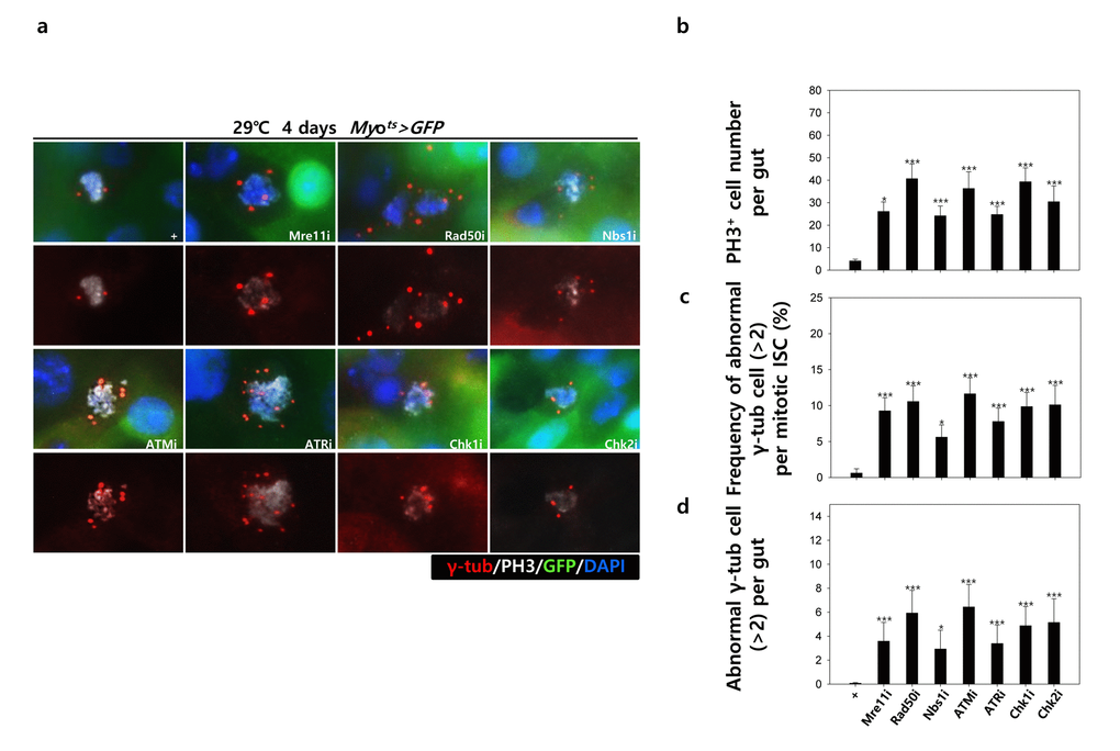
Figure 3B.EC-specific knockdown of DNA damage response (DDR)-related factors causes an increase in the age-related phenotypes of ISCs. EC-specific knockdown of DDR-related factors cause centrosome amplification in ISCs. Flies carrying Myots>GFP, Myots>GFP+Mre11i, Myots>GFP+Rad50i, Myots>GFP+Nbs1i, Myots>GFP+ATMi, Myots>GFP+ATRi, Myots>GFP+Chk1i, or Myots>GFP+Chk2i genotypes were cultured at 29 °C for 4 days. (a) The guts of flies were dissected and labeled with anti-GFP (green), anti-γ-tubulin (red), and anti-PH3 (white) antibodies and DAPI (blue). Original magnification is 400×. (b-d) Increased number of mitotic ISCs with supernumerary centrosomes (>2) in the guts of Myots>GFP, Myots>GFP+Mre11i, Myots>GFP+Rad50i, Myots>GFP+Nbs1i, Myots>GFP+ATMi, Myots>GFP+ATRi, Myots>GFP+Chk1i, or Myots>GFP+Chk2i flies. (b) EC-specific knockdown of Mre11, Rad50, Nbs1, ATM, ATR, Chk1, or Chk2 cause the increase of mitotic ISCs in the midguts. (c) Frequency of abnormal γ-tubulin cell per mitotic ISC. (d) Number of abnormal γ-tubulin cell per midgut. Three-day-old females were shifted to 29 °C for 4 days and dissected guts were immunostained with anti-GFP (green), anti-γ-tubulin (red), and anti-PH3 (white) antibodies and DAPI (blue). The centrosome numbers were counted in the PH3+ cells of these guts. Data (mean±SE) in Myots>GFP, Myots>GFP+Mre11i, Myots>GFP+Rad50i, Myots>GFP+Nbs1i, Myots>GFP+ATMi, Myots>GFP+ATRi, Myots>GFP+Chk1i, or Myots>GFP+Chk2i flies were collated from 61, 449, 557, 412, 560, 447, 687, and 349 mitotic cells of 15, 15, 11, 15, 13, 16, 15, and 9 guts, respectively. p-values were calculated using student’s t-test. *p<0.001, ***p<0.0001 compared to that of the Myots>GFP flies.