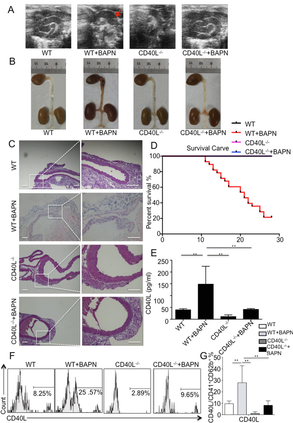
Figure 1.BAPN administration-induced TAD formation is inhibited by genetic depletion of CD40L. (A) Echography showed aortic arch dilation. (B) Representative images showed autopsy features of isolated mouse aorta after feeding with BAPN or saline for 28 days; arrow, location of TAD. (C) Haematixylin and eosin (H&E) staining showed significant dissected intima in WT+BAPN mice, while flat intima could be seen in other 3 groups. (D) Survival curve of WT (n=12), WT+BAPN (n=27), CD40L-/- (n=12) and CD40L-/-+BAPN (n=17) mice. (E) ELISA of sCD40L showed that circulating levels of CD40L are elevated in plasma from WT+BAPN mice. (F) Representative flow cytometry analysis showed percentage of CD41+ CD62b+ CD40L+ platelets divided by total CD41+ CD62b+ platelets in mice plasma. (G) Statistical data analysis from four separate experiments. Data were expressed as percentage ± SD of the CD41+ CD62b+ CD40L+ platelets subpopulations in total CD41+ CD62b+ platelets counted. **P < 0.01 versus WT, CD40L-/- and CD40L-/-+BAPN mice.