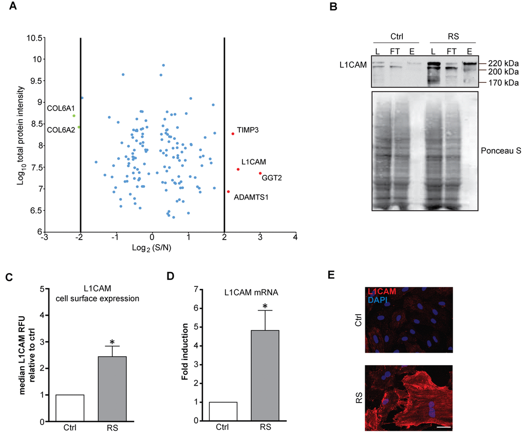
Figure 1.L1CAM is enriched on the surface of replicatively senescent fibroblasts. (A) Proteomic analysis of the surface of senescent cells. Down- (green dots < four fold) and up-regulated (red dots > four fold) proteins in senescent cells. (B) Cell surface proteins of proliferating (Ctrl) and replicatively senescent (RS) BJ fibroblasts were modified by biotin and captured on a streptavidin column (see Material and Methods for details), and fractions were validated for the presence of L1CAM by Western blotting. L – load (total protein); FT – flow through (non-biotinylated protein fraction), E – elution (biotinylated protein fraction). Ponceau S staining is shown to demonstrate protein loading. (C) FACS analysis of the surface level of L1CAM in BJ fibroblasts. (D) mRNA level of L1CAM normalized to GAPDH in replicatively senescent BJ cells. (E) Live cell immunofluorescence detection of L1CAM in proliferating (Ctrl) and replicatively senescent (RS) BJ (upper panel) and MRC5 (lower panel) fibroblasts. Scale bar, 50 μm. All experiments were performed in biological triplicates. For statistics, two-tailed Student’s t-test was used: p ˂ 0.05 (*); p ˂ 0.01 (**); p ˂ 0.001 (***).