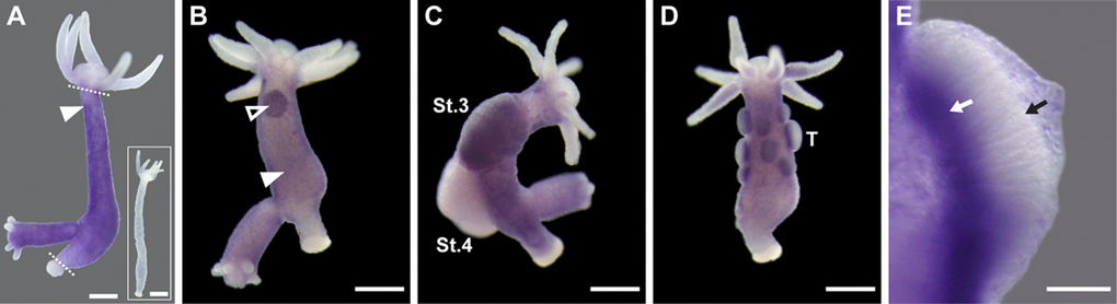
Figure 3.HyLMN is strongly expressed in the proliferating stem cells, but absent from the differentiated cells. (A) Whole-mount in situ hybridization with a DIG-labeled antisense probe specific for hyLMN reveals that the expression of hyLMN mRNA is restricted to the stem cells compartment (marked out by two dashed lines), with highest signal observed in the interstitial cells (arrowhead). Hybridization with a sense probe (inset) gives no signal. (B) In a sexually-induced polyp, hyLMN mRNA is expressed at elevated levels in single interstitial cells (white arrowhead) and clusters of precursor cells (empty arrowhead) typical for early gonad formation. (C) In a female polyp, a strong hyLMN signal is detected in the early gonad on stage 3 of oogenesis (St.3) and absent in later oogenesis stages (St.4). (D-E) In a male polyp, strongest hyLMN expression is observed in the basis of testes (T on D; white arrow on E), where mitotically dividing precursor cells are located. In the apical zone of the testis no signal can be detected (black arrow on E), indicating an absence of the hyLMN transcript in post-meiotic cells. Scale bar: 300 μm (A-D), 50 μm (E).