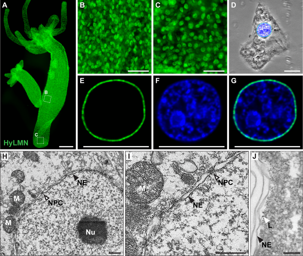
Figure 4.HyLMN protein is present in the nuclei of every Hydra cell and forms a nuclear lamina. (A-C) Immunostaining with anti-HyLMN antibodies reveals HyLMN protein in the nuclei of all cells in the polyp (A), including the stem-cell compartment (B) and the foot, made of differentiated cells (C). (D-G) HyLMN protein forms a thin layer - the lamina, surrounding the chromatin. Immunodetection of Lamin (green), DNA (blue), merged with phase contrast (on D). (H, I) Transmission electron microscopy reveals a typical organization of the nucleus in an epithelial cell of Hydra. Nuclear envelope (NE) consists of two membranes with incorporated nuclear pore complexes (NPC). The chromatin and a conspicuous nucleolus (Nu) are found within the nuclear envelope. Several mitochondria (M) are located close to the outer nuclear membrane. (J) Electron microscopy immunolocalization of the HyLMN protein shows that the lamina (L), labeled by the 6 nm gold particles, lays beneath the inner membrane of the nuclear envelope (NE). Scale bar: 300 μm (A), 50 μm (B-C), 10 μm (D-G), 500 nm (H-I), 100 nm (J).