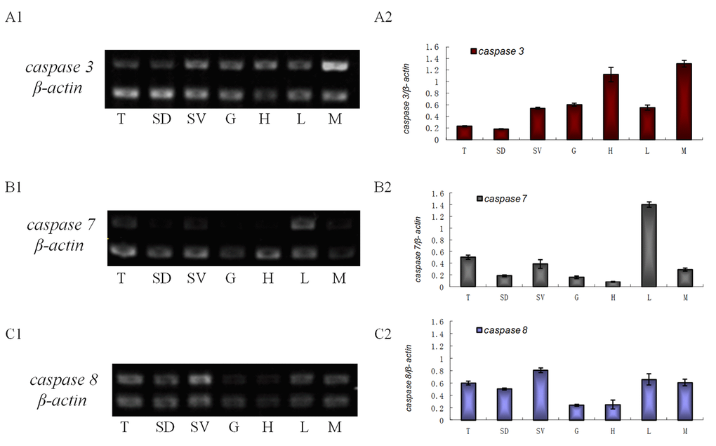
Figure 1.The transcriptional level of es-caspase 3/ es-caspase 7/ es-caspase 8 in different tissues. Seven tissues were dissected from E. sinensis: testis (T), seminiferous duct (SD), seminal vesicle (SV), gill (G), heart (H), hepatopancreas (L) and muscle (M). Histograms in the right were constructed from the agarose gel data in the left. (A1-A2) The semi-quantitative RT-PCR results of es-caspase 3. Higher es-caspase 3 was expressed in M and H. (B1-B2) The semi-quantitative RT-PCR results of es-caspase 7. Higher es-caspase 7 was expressed in L and T. (C1-C2) The semi-quantitative RT-PCR results of es-caspase 8. The reproductive system (T, SD and SV) showed higher expression of es-caspase 8. β-actin was used as the control. Data are the means of three independent experiments.