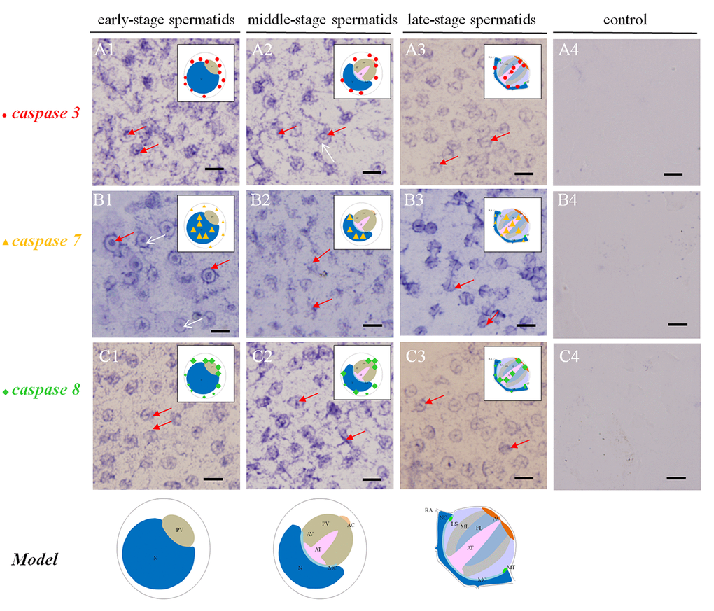
Figure 2.Temporal and spatial orientation of es-caspase3/ es-caspase7/ es-caspase8 during spermiogenesis in E. sinensis. The blue signals in each group were performed by ISH. N: nucleus, PG: proacrosomal granule, PV: proacrosomal vesicle, AT: acrosome tube, AC: acrosome cap, AV: acrosome vesicle, FL: fibrous layer, ML: middle layer, LS: lamellar structure, MC: membrane complex, RA: radical arm, NC: nuclear cap, MT: mitochondria. (A1-A3) The expression pattern of es-caspase 3 at various testis stages. In the early spermatids, es-caspase 3 was distributed in one pole of the cytoplasm and the inner edge of plasma membrane. Signals was decreased in middle-spermatids, and es-caspase 3 was distributed in the AT and AC finally. (B1-B3) The expression pattern of es-caspase 7 during spermiogenesis. Signals were discovered in the nucleus throughout the process. In the mature spermatids, es-caspase 7 was expressed in the AT, FL, ML and nucleus. (C1-C3) The distribution of es-caspase 8 in spermatids. The variation tendency was similar with es-caspase 3. The typical spermatid model was in the upper right corner of each panel. (A4-B4-C4) The control group. Bars=5 um.