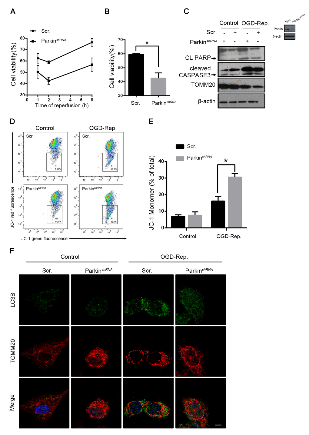
Figure 5.Parkin protected L02 cells from OGD-Rep. induced injury. L02 cells were transfected with Parkin shRNA (Parkin shRNA) or control shRNA (Scr.). Transfected L02 cells were subjected to OGD for 24 h, followed by recovery in normal cell culture medium and oxygen for reperfusion. (A) Cell viability was detected by CCK8 at 1, 2 and 6 h after reperfusion. (B) Quantification of cells viability at 2 h after reperfusion from indicated cells. (C) At 2 h after reperfusion, the whole cells lysate was collected. Cleaved PARP (CL PARP), cleaved CASPASE-3 and TOMM20 protein levels were determined by western blot analysis. (D) Measurement of mitochondrial membrane potential by JC-1 flow cytometry at 2 h after reperfusion. (E) Quantification of mitochondrial membrane potential loss. (F) LC3B (green) and the mitochondrial marker TOMM20 (red) were stained by immunofluorescence and the images were taken by confocal microscopy after 2 h of reperfusion. The data are expressed as mean ± SD. Statistical comparisons were performed with t-test. *P < 0.05 vs. the indicated group. Scale bar: 10μm.