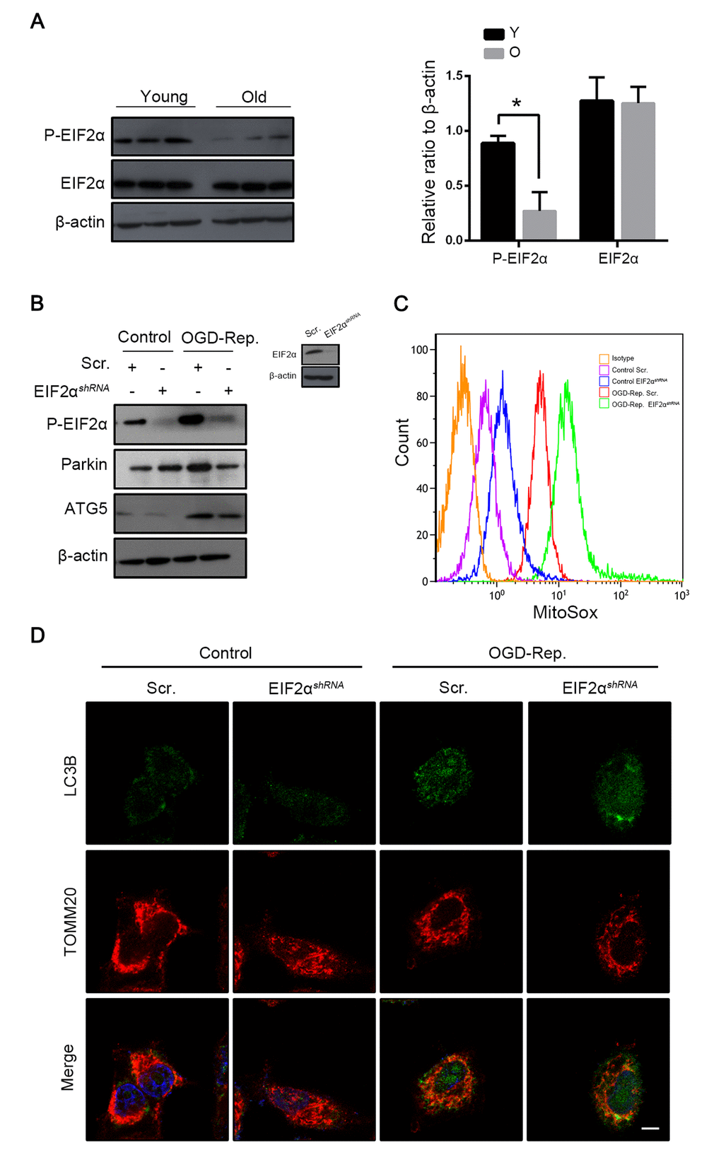
Figure 6.Phosphorylation of EIF2α induced mitophagy in L02 cells after OGD-Rep. with increased parkin expression. Mice of different age were treated as indicated (Y for young mice, O for old mice). (A) The EIF2α and phosphorylated EIF2α protein levels were determined by western blot analysis from the indicated groups. L02 cells were transfected with EIF2α shRNA (EIF2αshRNA) or control shRNA (Scr.). Transfected L02 cells were subjected to OGD for 24 h, followed by recovery in normal cell culture medium and oxygen for 2 h. (B) The whole cells lysate was collected and phosphorylated EIF2α, Parkin and Atg5 protein levels were determined by western blot analysis. (C) Mitochondria reactive oxygen species generation by was detected by MitoSOXred flow cytometry. (D) LC3B (green) and the mitochondrial marker TOMM20 (red) were stained by immunofluorescence. and the images were taken by confocal microscopy after 2 h of reperfusion. The data are expressed as mean ± SD. Statistical comparisons were performed with t-test. *P < 0.05 vs. the indicated group. Scale bar: 10μm.