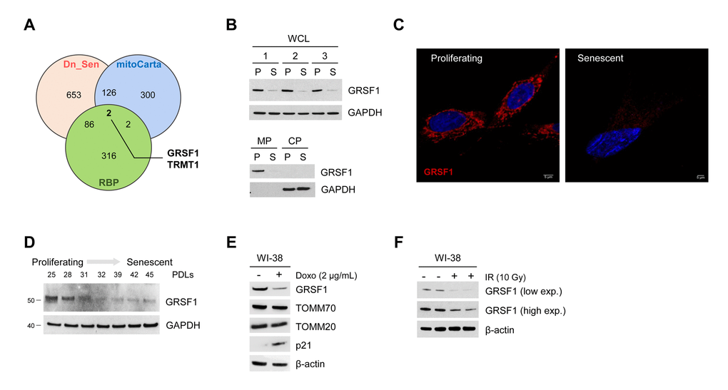
Figure 1.GRSF1 levels decline with senescence. (A) Venn diagram showing the overlap of mitochondrial RNA-binding proteins (overlap between mitoCarta and RBPs) that were significantly downregulated during replicative senescence (Dn_Sen). (B) GRSF1 protein levels were assessed in proliferating (P) and senescent (S) WI-38 fibroblasts in both WCL and in membrane (MP) and cytosolic (CP) fractions. (C) Representative image of GRSF1 immunofluorescence in P (population doubling level 23, PDL23) and S (PDL 58) WI-38 fibroblasts. (D) WI-38 cells of increasing PDLs were collected, and the levels of GRSF1 were assessed by Western blot analysis. (E,F) WI-38 fibroblasts (PDL23) were rendered senescent via different methods: by treatment with Doxorubicin (2 µg/mL) for 24 h followed by culture for an additional 7 days (E) and by exposure to 10 Gy of ionizing radiation (IR) followed by culture for 10 days (F). The indicated proteins were assessed by Western blot analysis.