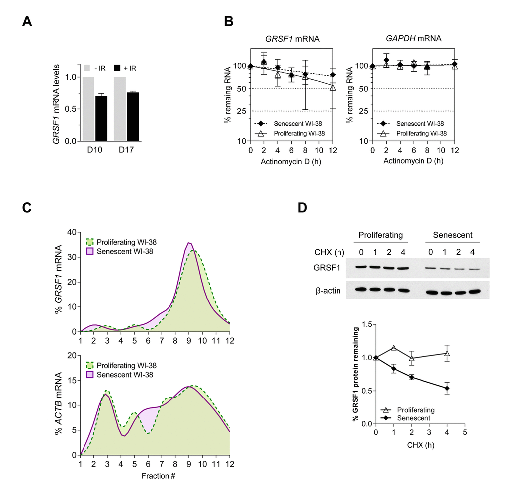
Figure 2.GRSF1 protein is unstable in senescent cells. (A) WI-38 fibroblasts were rendered senescent by exposure to 10 Gy of ionizing radiation (IR) followed by incubation for 10 days. Total RNA was then extracted and RT-qPCR analysis was used to measure GRSF1 mRNA levels and 18S rRNA levels (for normalization). (B) The stability of GRSF1 mRNA was studied by incubating proliferating (PDL23) and senescent (PDL55) WI-38 fibroblasts with actinomycin D and measuring the rate of clearance of GRSF1 mRNA (and GAPDH mRNA, a stable mRNA encoding housekeeping protein GAPDH) by RT-qPCR analysis; mRNA levels were normalized to 18S rRNA levels, quantified in the same samples. (C) GRSF1 translation was compared between proliferating (PDL23) and senescent (PDL55) WI-38 fibroblasts by fractionating polysomes and assessing throughout the sucrose gradients the distribution of GRSF1 mRNA and ACTB mRNA [encoding the control protein ACTB (β-Actin) and chosen because it is not generally subject to translational control]. Fractions 1 and 2 were devoid of ribosome particles, fractions 3-5 contained ribosomal subunits and monosomes, fractions 6-8 contained low-molecular weight polysomes, and fractions 9-12 contained high-molecular-weight polysomes (not shown). (D) The relative stability of GRSF1 protein in proliferating and senescent WI-38 fibroblasts was measured following incubation with 100 µg/mL of cycloheximide to block de novo protein synthesis. Lysates were prepared at the times shown and Western blot analysis was used to assess the levels of GRSF1 and β-Actin; after quantification by densitometry, the relative signal intensities were plotted. Data in (A,D) represent the means and S.D. from two independent experiments. Data in (B) represent the means and S.D. from three independent experiments.