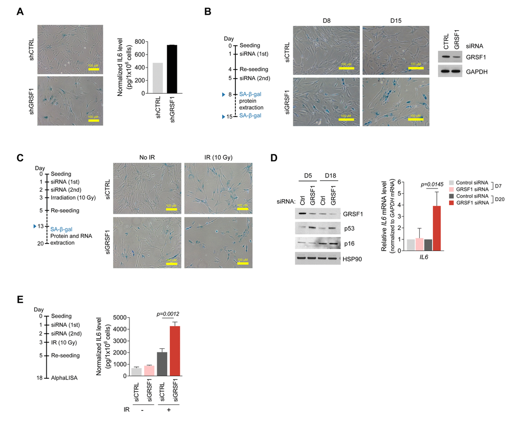
Figure 4.Silencing GRSF1 promotes senescence and IL6 production. (A) Three weeks after silencing GRSF1 by infection with shGRSF1- or shCTRL-expressing lentiviruses, SA-β-gal activity was assessed by light microscopy (left) and secreted IL6 was quantified by ELISA (right). (B) Pre-senescent WI-38 fibroblasts were transfected with siGRSF1 or siCTRL; SA-β-gal activity (micrographs) was assessed at day 8 (D8) and D15 after transfection. At D8, GRSF1 and GAPDH levels in WCL were assessed by Western blot analysis (right). (C,D) WI-38 cells transfected with siGRSF1 or siCTRL were irradiated (IR, 10 Gy) and subsequently cultured for 10 days; whereupon SA-β-gal activity was assessed (C); proteins in WCL prepared at D5 and D18 were assessed by Western blot analysis (D, left). RNA was extracted at D7 and D20, and IL6 mRNA levels were quantified by RT-qPCR analysis (D, right). (E) After transfection with siGRSF1 or siCTRL and irradiation, WI-38 cells were cultured in normal medium for 14 days and in serum-free condition for 24 h (left). CM was then collected, and the secreted IL6 was assayed by AlphaLISA (right). Data in (D,E) represent the means ±S.D. from three independent experiments.