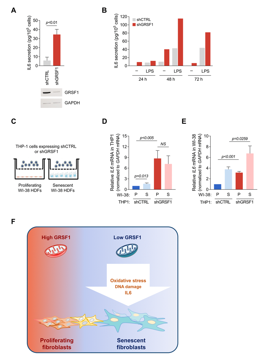
Figure 5.Loss of GRSF1 increases the paracrine actions of IL6. (A) THP-1 monocytes expressing lentiviral shGRSF1 or shCTRL were maintained in normal culture medium (containing 10% FBS), and the amount of IL6 secreted into the CM was measured by ELISA. (B) Human monocyte THP-1 cells were infected with shGRSF1 or shCTRL lentiviruses to produce different steady-state levels of GRSF1 constitutively. The levels of IL6 secretion by these cells was measured by ELISA; treatments with LPS (50 ng/mL) for 24, 48, and 72 h were included as positive controls for IL6 production. (C-E) Schematic of the co-culture setup. Proliferating (PDL20-25) or senescent (PDL55-60) WI-38 fibroblasts were plated in the lower chamber, and THP-1 cells constitutively expressing different levels of GRSF1, as explained in (A), were plated in the upper chamber. The chambers were separated by a selectively permeable membrane with 0.4 µm-diameter pores (C). Lentiviral shRNA-expressing THP-1 cells were co-cultured with either proliferating or senescent WI-38 fibroblasts for 48 h, and the levels of IL6 mRNA in THP-1 cells (D) and WI-38 cells (E) were quantified by RT-qPCR analysis. Data in A,D,E represent the means ±S.D. from three independent experiments. (F) Model: we propose that loss of GRSF1 contributes to cellular senescence characterized by oxidative stress, DNA damage, growth suppression, and IL6 production.