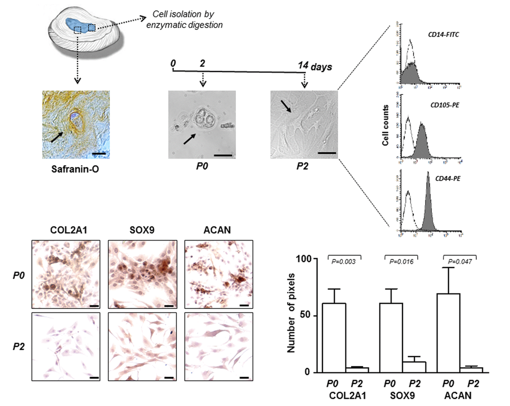
Figure 1.IVD cells: culturing and characterization. Representative optical photomicrographs showing morphology of the cells (indicated by the arrows) in the IVD tissue stained with Safranin-O, and at P0 and P2 passages in culture. P2 cells were characterized by flow cytometry for the expression of CD14 haematopoietic marker, and CD105 and CD44 mesenchymal markers. Flow cytometric analysis of a representative case is reported; open histograms represent the isotype control antibody, gray histograms represent anti-CD14, -CD105 and -CD44 antibodies. X-axis, fluorescent channel; Y-axis, number of events. Representative optical photomicrographs of COL2A1, SOX9 and ACAN immunostaining performed in P0 and P2 cells are reported. Protein expression levels were quantified by densitometric analysis of immunocytochemical pictures using ImageJ software and expressed as means of pixels per one hundred cells ±SD (P0 group, n = 6; P2 group, n = 6). Exact P-values are reported. Scale bars: 20 μm.