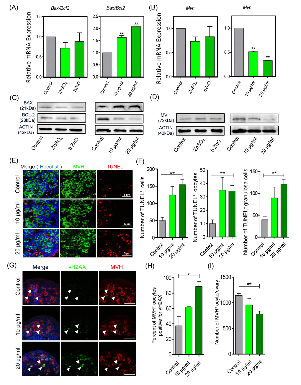
Figure 2.Apoptotic and DNA damage markers in nZnO treated fetal oocytes in vitro. (A-D) Representative q-RT-PCR and WB analyses of Bax, Bcl-2 and Mvh of ovarian tissues cultured for 6 days; Actin or Mvh was used as housekeeping gene and loading control, respectively. (E) TUNEL-staining of the ovarian tissues after 6 days of culture with nZnO. (F) Number of TUNEL-positive total cells, TUNEL-positive oocytes and TUNEL-positive granulosa cells per section. (G) Co-immunostaining of DNA damage marker γH2AX (green) and germ cell marker MVH (red) in ovary sections after 6 day nZnO exposure, Hoechst 33342 (blue) was used for nuclei staining. (H) Percentage of MVH positive oocytes also stained for γH2AX. (I) Number of MVH positive oocytes/ovary.