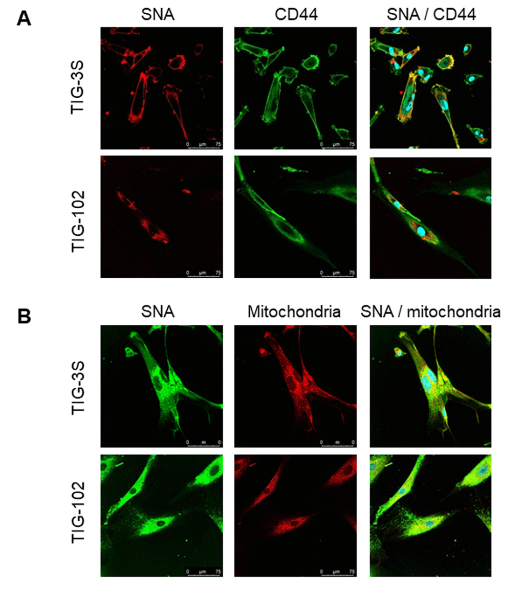
Figure 5.Localization of sialylated glycoproteins in TIG cells. (A) TIG-3S (PDL 38; top) and TIG-102 (PDL 46; bottom) stained with SNA (red; left panel), FITC-conjugated membrane marker (CD44, green; middle panel) and the overlay image (right panel). (B) TIG-3S (PDL 52; left) and TIG-102 (PDL 46; right) stained with FITC-conjugated SNA (green; left panel), an intracellular marker (mitochondria, red; middle panel), and the overlay image (right panel). Blue staining represents the nucleus.