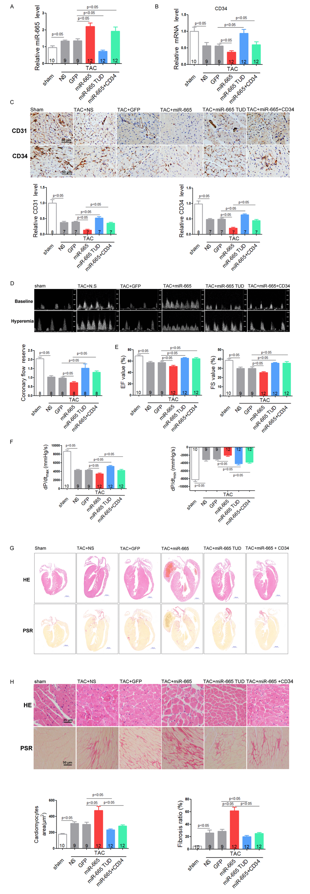
Figure 6.Inhibition of miR-665 or re-expression of CD34 improves cardiac dysfunction via angiogenesis in TAC mice. (A) Relative cardiac expression of miR-665 detected by real-time PCR. (B) Relative CD34 expression level in heart detected by real-time PCR. (C) Representative images of immunohistochemical staining for CD31 and CD34 in heart tissues. (D) Representative images of Pulsed-wave Doppler of coronary artery at baseline or under hyperemic conditions induced by inhalation of 1% or 2.5% isoflurane, respectively. The coronary flow reserve is calculated as the ratio of hyperemic peak diastolic flow velocity to baseline peak diastolic flow velocity. (E) Echocardiographic detection in treated mice. (F) Hemodynamic analysis measured by Millar cardiac catheter system in treated mice. (G) Gross morphology by hematoxylin and eosin (H&E) staining and picrosirius red staining of hearts from treated mice. (H) Histological analysis of surface area of cardiomyocytes by H&E staining and collagen deposition in heart by picrosirius red staining. The numbers of mice tested are showed in the bars. Data are expressed as mean ± SEM.