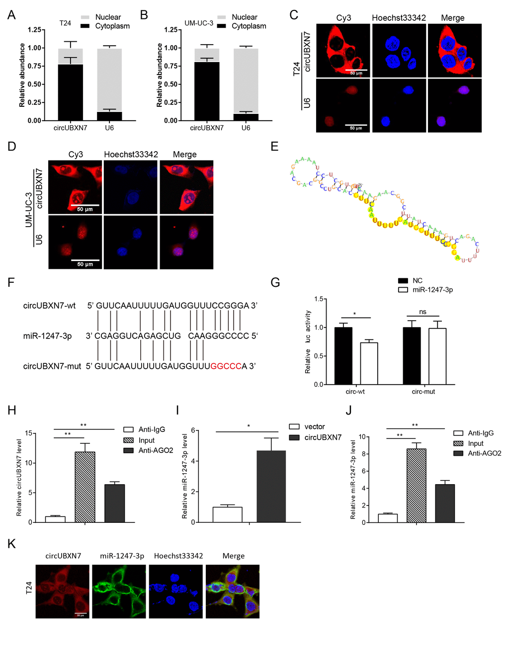
Figure 4.CircUBXN7 directly bound to miR-1247-3p. (A and B) Subcellular distribution of circUBXN7 was determined by nuclear mass separation assay. (C and D) FISH assay for investigating the subcellular localization of circUBXN7 in T24 and UM-UC-3 cells. U6 was used as a nuclear control. Scale bar, 50μm. (E) The secondary structure of circUBXN7 that possibly binds to miR-1247-3p was predicted by RegRNA 2.0. The yellow region indicates the predicted motif structure. (F) The potential binding sites between circUBXN7 and miR-1247-3p were predicted by RNAhybrid. The red part represents the mutated base. (G) miR-1247-3p reduced the luciferase activity of circUBXN7 in 293T cells detected by dual-luciferase activity assay. (H) Anti-AGO2 RIP assay pulled down more circUBXN7 than in the anti-IgG group. (I) Relative miR-1247-3p levels immunoprecipitated by AGO2 in circUBXN7 overexpressing or control cells. (J) Relative miR-1247-3p levels immunoprecipitated by AGO2 or IgG in circUBXN7 overexpressing cells. (K) Colocalization of circUBXN7 and miR-1247-3p was detected in T24 cells by FISH assay. Scale bar, 50μm. *P<0.05, **P<0.01.