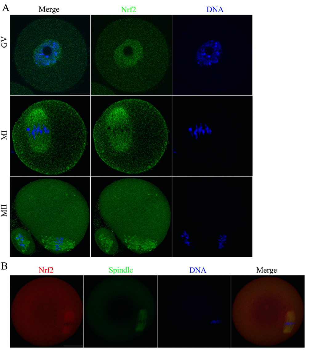
Figure 2.Cellular distribution of Nrf2 during oocyte meiosis. (A) Immunofluorescent staining was employed to show the subcellular localization of Nrf2. Green, Nrf2; Blue, chromatin. A total of 30 oocytes were examined for each group. Scale bar, 20 μm. (B) Immunofluorescent staining for co-localization of α-tubulin with Nrf2 in mouse oocytes. This shows the co-localization of Nrf2 with spindles in mouse oocyte. Red, Nrf2; green, tubulin; blue, chromatin. Scale bar, 20 μm.