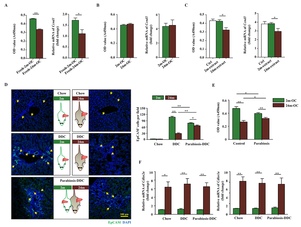
Figure 2.The altered microenvironment in aged mice affects the activation of OCs. Young (2m) and aged (24m) mice were fed with DDC diet for 3 weeks. (A) Freshly isolated OCs from young (Fresh-2m-OC) and aged (Fresh-24m-OC) DDC-fed mice were cultured on type I collagen coated 96-well plates, 3 days later, CCK-8 test was performed (left panel, n=6, ** p < 0.01). Quantitative Real-time PCR analysis of Ccnd1 in 2m-OC and 24m-OC (right panel, n=6, * p < 0.05). (B) Freshly isolated OCs were passaged for 6 times, then CCK-8 test (left panel, n=6) and quantitative Real-time PCR analysis of Ccnd1 (right panel, n=6) were performed. (C) Freshly isolated OCs were passaged for 6 times, then were cultured in normal medium (Ctrl), with liver extract from young mice (2m-extract) and with liver extract from aged mice (24m-extract). CCK-8 test (left panel, n=6, * p < 0.05) and quantitative Real-time PCR analysis of Ccnd1 (right panel, n=6, * p < 0.05) were performed. (D) Young and aged mice were joined in parabiotic pairs for 3 weeks with DDC diet. Immunofluorescence staining for EpCAM+ cells (green) was performed. Quantification of EpCAM+ cells was shown (n=6, * p < 0.05, ** p < 0.01). (E) CCK-8 test was performed in the OCs freshly isolated from the parabiotic pair (Parabiosis) or individuals as controls (Control) (n=6, * p < 0.05, ** p < 0.01). (F) Young and aged mice were joined in parabiotic pairs and kept for 3 weeks under DDC diet. The transcript levels of Cdkn2a, and Cdkn1a in the liver tissues were measured by quantitative real-time PCR (n=5, * p < 0.05, ** p < 0.01).