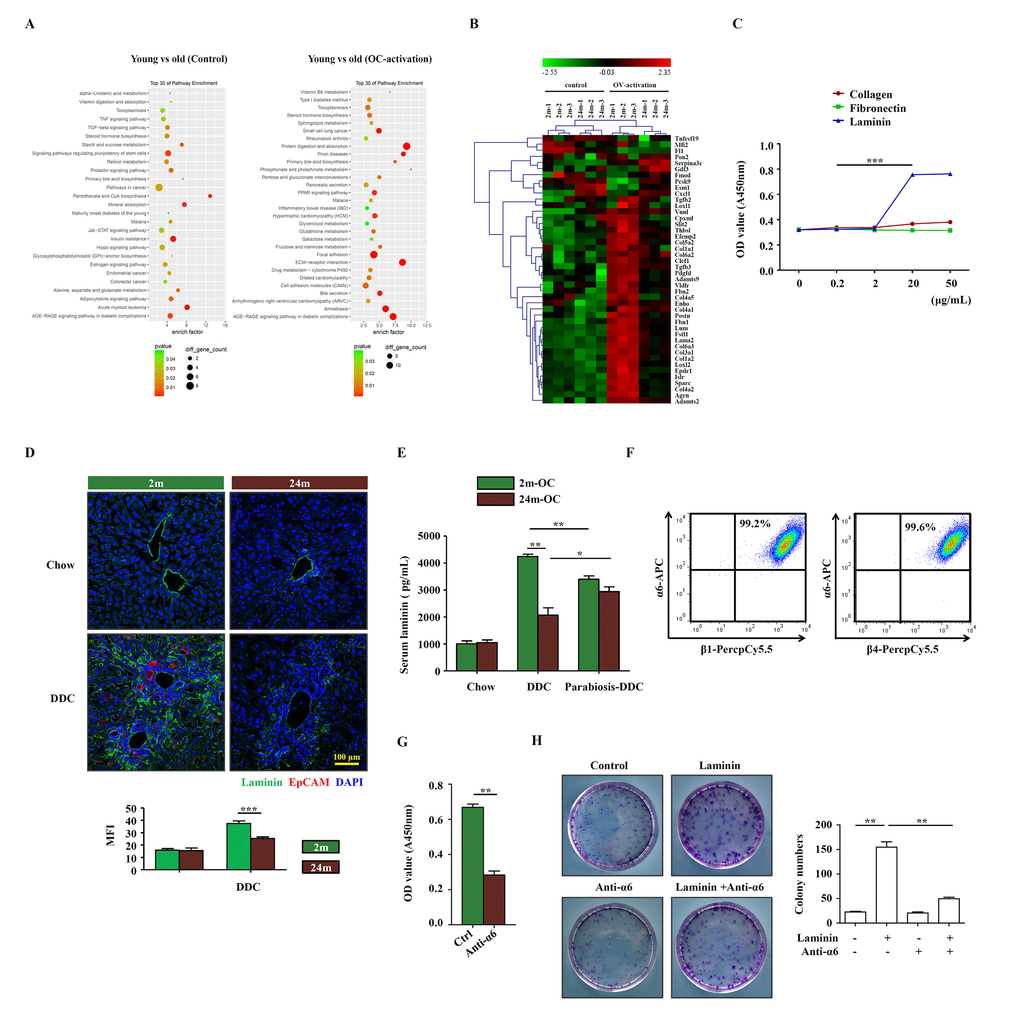
Figure 3.Laminin supports OC proliferation via integrin signaling pathway. (A) Microarray analysis of young and aged control/OC-activated mice liver was performed. Result of pathway enrichment analysis was shown. (B) Heatmap of 44 genes with significant expression difference (fold change > 5, p < 0.01) between young (2m) and aged (24m) control/OC-activated mice liver (3 samples for each group). (C) Different dose of collagen, fibronectin and laminin were added to the OC culture medium and the proliferation of OCs was analyzed by CCK-8 (versus 0.2 mg/mL, n=5, *** p < 0.001). (D) Immunofluorescence staining for laminin (green) and EpCAM (red) in DDC-fed (DDC) versus chow controls (Chow). MFI of laminin was quantified (n=6, *** p < 0.001). (E) Young and aged mice were joined in parabiotic pairs and kept for 3 weeks under DDC diet. The level of serum laminin was detected by ELISA (n=5, * p < 0.05, ** p < 0.01). (F) Flow cytometry showed the expression of α6, β1 and β4 integrins on OCs. (G) CCK-8 test showed that the proliferation of OCs treated with anti-α6 integrin antibody (n=6, ** p < 0.01). (H) Colony-forming assay of freshly isolated EpCAM+ cells from DDC diet with/without laminin (20 μg/mL) and anti-α6 integrin antibody (2 μg/mL). Pictures were representative wells of each condition. Colony number was quantified (n=6, ** p < 0.01).