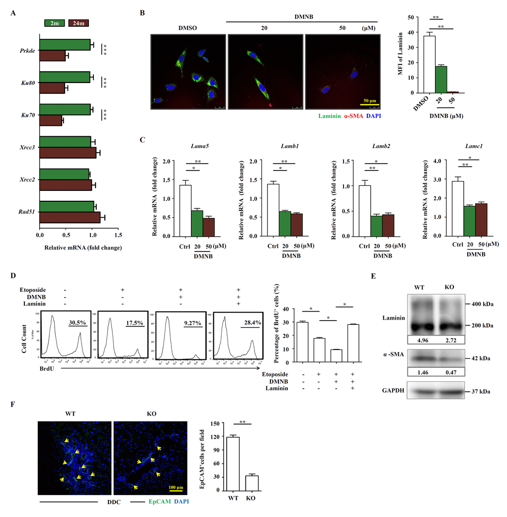
Figure 6.The OC-supporting function of HSCs relies on DNA-PK pathway. (A) Quantitative Real-time PCR analysis of HR and NEHJ components in HSCs freshly isolated from young (2m) and aged (24m) mice with DDC diet (n=4, *** p < 0.001). (B) The expression of α-SMA (red) and laminin (green) in etoposide (0.1 μM) and DMNB treated JS1 cells was detected by confocal. MFI of laminin was quantified (n=5, ** p < 0.01). (C) Quantitative Real-time PCR showed that the expression of laminin isoforms in etoposide (0.1 μM) and DMNB treated JS1 cells (n=6, * p < 0.05, ** p <0.01). (D) Proliferation of OCs co-cultured with JS1 cells pretreated with etoposide, DMNB or laminin was compared by BrdU analysis. BrdU+ cell percentage was quantified (n=4, * p < 0.05). (E) Western blot analysis of indicated proteins in HSCs isolated from wild type (WT) and Ku80 knockout (KO) mice fed with DDC. (F) Immunofluorescence staining for EpCAM+ cells (green) in DDC-fed wild type (WT) versus Ku80 knockout mice (KO). Quantification of EpCAM+ cells was shown (n=5, ** p <0.01).